
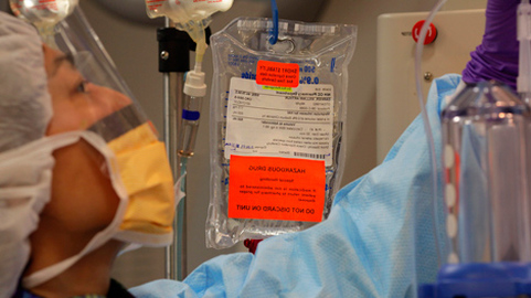
Today brings news that scientists at the US National Toxicology Program found that exposure to cell phone radiofrequencies increased brain tumours in male (but not female) rats. Here is a CTV.ca blog from 2010 examining the evidence that cell phones cause brain tumours in humans.
Cellphone safety study: Missing the answer by adhering to a flawed principle
May 17, 2010 08:50 by Dr. Lorne Brandes
As most people know, the safety of cellphones has been a hot topic of debate since several published reports suggested a possible increase in ipsilateral (same-side) brain tumours (malignant gliomas and benign meningiomas) among heavy users.
Although health authorities cautioned that the data from these studies were insufficient to provide a definitive answer, concerned governments from Ontario to India were quick to give advice: children, whose brains might be especially vulnerable to the effects of radiofrequency (RF) emissions, should be discouraged from using cellphones, except in an emergency.
Unfortunately, with today’s publication of the highly-awaited Interphone Study Group report, those hoping for that “definitive” answer are certain to be confused. But should they?
This largest-to-date study, conducted between 2000 and 2004, involved 13 countries, including Canada. Carried out under the auspices of the World Health Organization’s International Agency for Research on Cancer (IARC), it involved 2,708 glioma patients, 2,409 meningioma patients, and 2,972 carefully matched healthy controls.
The subjects, all between the ages of 30 and 59, were interviewed in person to determine the frequency and cumulative call time (total hours) of cellphone use (or non-use) over several time spans: 1 – 1.9 years; 2 -4 years; 5 – 9 years; 10 or more years.
The findings? As compared to “never-users”, there was an overall 20 to 30% decreased risk of glioma and meningioma tumours in those who used cell phones for 1 -9 years; even after 10 or more years of use, a 2% decreased risk of glioma and a 17% decreased risk of meningioma was found.
When cumulative call time was compared among ever- and never-users, once again a 20 to 30% decreased risk of tumours was observed among users who accumulated less than 1,640 hours over 1 to 10 years.
However, once cumulative call times went above 1,640 hours for 10 or more years of use (approximately 30 minutes daily), a 15% increase in meningiomas and a 40% increase in gliomas was observed. Perhaps tellingly, the rate of gliomas was a whopping 377% higher in those who accumulated more than 1640 hours over just 1 – 4 years (i.e., ranging from 1.1 – 4.5 hours of daily use). Moreover, as suggested in the earlier studies, their location favored the ipsilateral temporal lobe (the part of the brain closest to the ear).
Despite the study’s findings (both “good” and “bad”), the committee of authors was extremely reluctant to draw any conclusion at all, stating that “[methodological] biases and error prevent a causal interpretation…” As a result, they called for yet further investigation on “the possible effects of long-term heavy use of mobile phones”, especially among adolescents and teenagers.
The reason is this: they were concerned that an excess of brain tumours was seen only at the highest level of use, with no evidence of a linear increase up to that point. Indeed, except at the highest level of use, there appeared to be a protective effect of cellphones against brain tumours -- something the authors dismissed as “implausible”… so implausible that, despite other studies showing a similar trend, the Interphone Study Group is “currently exploring the possibility of correcting the [risk] estimates mathematically”. And with that statement, if any bias exists, it appears to be more in the thinking of the investigators than as a result of the design of the study.
Why? For decades, health officials, toxicologists and regulators, including those associated with the IARC, have only recognized the validity of what is called a “linear (straight line) dose-response” effect. But, based on hundreds upon hundreds of published examples of chemical, hormonal and physical agents, whose dose responses are “hormetic” (derived from the word, hormone, meaning differing effects at low and high levels of exposure), many prominent scientists, chief among them, U. of Massachusetts professor, Dr. Edward Calabrese, believe that the hormetic model, rather than the linear model, may more accurately represent the biological effect, especially at lower levels of exposure, of many agents on human health.
One example is the “J-shaped” curve that applies to alcohol; as compared to non-drinkers, decreased cardiovascular-related mortality is observed in populations that consume up to 2 ounces per day; exceeding that amount results in higher mortality (including cancer) that increases the more that one drinks.
Furthermore, although there is evidence of benefit when plants and animals are exposed to low-level terrestrial and extra-terrestrial radiation, the IARC disregards studies of low-dose radiation effects, believing instead that there is no safe lower threshold of human exposure to radiation. That thinking likely extends to RF emissions from cell phones.
So what is the truth about cell phones and the risk of brain tumours? I believe that the answer lies in the hormetic model proposed by Dr. Calabrese and his colleagues. A “J-shaped” dose response (decreased risk of brain tumours with low to moderate use; increased risk with excessive use) is highly consistent with the findings of the Interphone Study Group. Sadly, the “linear thinkers” will say otherwise. Officially, nothing will have been resolved and stiff-necked adherence to a seriously flawed risk model will have obscured highly pertinent findings.
May 24th, 2016
Important new guidelines for doctors and patients on treatment for advanced cancer
April 11, 2012 07:46 by Dr. Lorne Brandes
As an oncologist, I can say without hesitation that nothing in my daily practice compares to the unhappy duty of breaking bad news. Telling a patient with progressing cancer that further chemotherapy treatments are unlikely to be effective signals the end of the hopeful phase of their journey and an inevitable transition to palliative care.
Such discussions are especially difficult when they involve younger patients. “You mean you’re giving up on me?” is the (usually) unasked question reflected in their facial expression. Yet, no matter how delicately and sympathetically the subject of “no further treatment” is broached, some patients shoot the messenger, blaming the doctor for failing them, and demanding a referral to another oncologist.
Sometimes, recommending that treatment be stopped can be made even more difficult by the knowledge that some colleagues continue to recommend treatment beyond the “norm,” even when there is no hope for a meaningful outcome.
Most often, this refusal to “throw in the towel” reflects a genuine concern that no stone be left unturned. But when is such an approach unreasonable and counter-productive? After all, chemotherapy has toxic side effects, and the quality of remaining life is an exceedingly important issue to patients coping with a terminal disease.
Moreover, a difficult situation is often made worse by the almost daily reporting by the media of new cancer “breakthroughs” that, in truth, are only in the test tube, or laboratory mouse, stage of development. “Why can’t you try that new treatment on me?” some will ask out of desperation.
Thankfully, new guidelines issued by the American Society of Clinical Oncology (ASCO), and published in this week’s edition of the Journal of Clinical Oncology (JCO), go a long way in identifying who should, and should not, be considered for further chemotherapy treatment when metastasized cancer progresses on therapy.
The ASCO guidelines are part of a broader program, called “Choosing Wisely,” first proposed in a 2010 commentary in the New England Journal of Medicine by Dr. Howard Brody, PhD, director of the Institute for the Medical Humanities and a professor of family medicine at the University of Texas. In his wide-ranging perspective, Dr. Brody challenged medical specialties to take a critical look at their fields and to each identify five practices that are commonly performed despite a lack of supporting evidence.
"At ASCO, we took this challenge to heart," said Lowell E. Schnipper, MD, lead author of the JCO article and chair of ASCO's Cost of Care Task Force. "By tackling the overuse of treatments and tests for some of the most common cancers, we hope to achieve substantial improvements in the quality of cancer care…The Top Five list is just the first step in an ongoing ASCO effort to help physicians and patients implement these recommendations."
"As oncologists, we have a responsibility to help ensure that all cancer care is high-value care. That means providing the highest quality of care to our patients, while avoiding treatments that have little or no proven benefit,” added Dr. Michael P. Link, president of ASCO.
Topping the list of five common [oncology] practices that lack supporting evidence is “unnecessary anticancer therapy, including chemotherapy, in patients with advanced solid-tumor cancers [such as breast cancer] who are unlikely to benefit.”
According to the ASCO committee, “Data have shown that a significant number of cancer patients receive chemotherapy in the last two weeks of life, even though such treatment generally does little to improve survival or quality of life, causes side effects and carries the unintended consequence of increasing costs. Such care may also postpone patients' access to palliative care, including hospice care.
“ASCO recommends that cancer-directed therapy not be used for solid tumor patients with the following characteristics: low performance status [i.e., bedridden 50% or more each day], no benefit from prior evidence-based interventions, not eligible for a clinical trial, and no strong evidence supporting the clinical value of further anti-cancer treatment.
“Because further treatment is unlikely to be effective in these patients, emphasis should be placed on palliative and supportive care, which can increase quality of life and, in some cases, extend survival.”
Exceptions were duly noted by the committee: previously untreated patients with a low performance status due to other (non-cancer) causes should still be considered for evidence-based chemotherapy, as should patients with progressive disease whose tumours have “molecular markers [identifiable gene mutations]” that respond to some form of drug therapy.
So what is the bottom line? The ASCO chemotherapy guidelines make eminent sense and are almost certain to set the standard of care for chemotherapy in advanced cancer.
Personally speaking, I plan to give a copy of the ASCO recommendations to each and every patient whose cancer progresses on chemotherapy so that they may better understand why continuing on with yet more treatment may not be in their best interest.
Do doctors spend too little time with patients?
May 23, 2012 09:13 by Dr. Lorne Brandes
If there is one topic about which almost every Canadian has an opinion, it is medical remuneration. While the debate usually centres around whether doctors’ high incomes, paid out of the public purse, are justified, two recent and unrelated events have raised the issue of how much time doctors should spend with patients to earn that income.
In the first instance, a Manitoba woman seeing her doctor about back pain was allegedly told she would have to make another appointment to discuss her concerns about chest pain. According to her husband, two weeks later the woman died of a heart attack in a hospital emergency ward. The husband has written a letter to the Manitoba College of Physicians and Surgeons asking for a full investigation.
Despite a paucity of any real facts surrounding this case, a raw nerve was exposed. The public outcry was both reflexive and negative.
Noting that people "can relate to someone who has felt short-changed after being rushed through a medical appointment," an editorial in the Winnipeg Free Press quickly got down to the nuts and bolts:
"The story of the woman who was not allowed to complain to her physician about chest pains -- one problem per visit, she was told -- seems apocryphal. It is, after all, the antithesis of good medicine. But it is not impossible, nor improbable because medicine is also a business and doctors charge fees for service on a per-visit basis."
The editorial went on to identify two contrasting sides of the issue:
- On one hand, "the Manitoba College of Physicians and Surgeons is getting increasingly worried about the practice followed by some doctors, often posted on office walls, that restricts clients to a ‘one problem per visit’ rule."
- On the other hand, "the Canadian Medical Association... suggests doctors tell clients there is a limit to the time and number of complaints that can be handled in a visit" that, in Manitoba, typically pays a family doctor $31.85 for a minimum of 10 minutes, with no maximum.
- "The [fee tariff schedule] must recognize that some patients and conditions will take much more time to diagnose. Doctors must be compensated reasonably for the time."
- "Doctors must be reminded that they are professionally bound to deliver good medicine; that involves listening respectfully to [patients] who cannot know, but might suspect, that a number of aches can be tied to an unsuspected and potentially serious health problem."
For example, in Manitoba, an eye surgeon currently receives $513.05 for a cataract operation, defined in the physician’s manual as "extraction of lens with insertion of intraocular implant—unilateral, with or without iridectomy". Historically, the calculation of the fee was based on assigning a "value unit" for each 15 minutes of operative time of $21.375 (or about $85.00 per hour).
Critics argue that, several years ago, when cataract surgery took 4 hours on average, eye surgeons were fairly compensated. However, as a result of rapid technological advances, the same procedure can now be completed in around 30 minutes, freeing up time for the doctor to perform additional cataract surgeries. Therefore, based on the "value unit" calculation, should not the fee for each procedure be significantly lower?
While one is tempted to agree that, based on standard business models, increased efficiency and output should result in lower cost, how does one compare the pricing of a commodity to the fee paid for the exacting skill and training (including learning and using the latest new techniques) of an ophthalmologist? Is it not apples and oranges?
Yet, the crux of the problem is that doctors have always been paid more for doing than thinking. And it is no secret that surgeons and other specialty groups have always been very proactive in protecting their procedural turf in any fee negotiations.
As a result, the much larger and less homogeneous group of family doctors, who serve as the primary gatekeepers of our health care system, have usually gotten the short end of the financial stick. As exemplified by "one complaint per visit", we all know where that can lead, and it’s not to a place we like.
At the end of the day, both aspects of doctors spending "too little time" must be resolved in ways that are fair and that address inequities in the fee schedule without sacrificing patient welfare.
While this may well require a realignment in doctors’ remuneration, any unilateral rollback of previously negotiated procedural fees, such as those taken by the government of Ontario Premier Dalton McGuinty, and now being considered by other provinces, should be condemned as politically short-sighted and destructive to future relations between governments and doctors.
My suggestion for a fix? In a system with finite dollars for medical remuneration, family doctors and internists should be paid significantly more to think. This should be counter-balanced, at least in part, by surgeons and radiologists voluntarily agreeing to accept a reasonable fee reduction for procedures that they can now perform more quickly and more often.
Crossing the Rubicon: Hailing a new era in cancer treatment
June 4, 2012 08:29 by Dr. Lorne Brandes
Unlike all too many previous occasions, this week’s annual meeting of the American Society of Clinical Oncology (ASCO) provided a palpable sense of real progress in the “war against cancer” as a virtual torrent of promising new clinical treatments emerged.
Primed by advanced publicity, much of it from drug companies , thousands of participants literally ran from auditorium to auditorium in Chicago’s cavernous McCormick Place to hear the results of “breakthrough” trials involving novel drugs and vaccines that, better than anything before them, effectively shrink a variety of hard-to-treat, advanced cancers.
And truth be told, in many cases the data lived up to the hype. As a result, it would not be an overstatement to suggest that, like Caesar in 49 BC, cancer treatment in 2012 crossed the Rubicon ; there will be no turning back!
One prime example is an exciting new drug, called T-DM1 , for advanced, aggressive (Her2-positive) breast cancer that I discussed in a blog last year. Co-developed by Roche and ImmunoGen, T-DM1 is a molecular hybrid, formed by linking the Her2 receptor antibody, trastuzumab ( Herceptin ), with emtansine (DM1), a derivative of a naturally-occurring toxic chemical called maytansine .
Herceptin alone benefits approximately 15% of women with advanced breast cancer; to be most effective, it needs to be administered with chemotherapy drugs, called taxanes, that produce a lot of unwanted side effects. Emtansine is too toxic for use in humans. However, when the two are linked up to form T-DM1, the result is a robustly-active drug with high specificity and a low incidence of side effects.
Why? Because, by specifically binding to the Her2 receptor, the Herceptin component of T-DM1 delivers the toxic emtansine directly into Her2-positive breast cancer cells, bypassing normal cells.
Early results in a phase 2 study were impressive; one-third of 112 women with Her2-positive breast cancer whose tumours had progressed on Herceptin, responded to T-DM1, some dramatically . Side effects were generally mild.
So, when Roche announced in May that “positive results” of its follow-up phase 3 study of T-DM1, involving almost 1000 women with metastatic Her2-positive breast cancer that had progressed on Herceptin/taxane treatment, would be presented at ASCO, it was not surprising that the company quickly got everyone’s attention.
And when the outcome of that prospective randomized trial was finally presented on Sunday, June 3rd, it did not disappoint.
Compared to women who received standard 2nd line treatment [an oral anti-Her2 drug, called lapatinib (Tykerb), combined with an oral chemotherapy drug, called capecitabine (Xeloda)], those who were treated with T-DM1 lived 3 months longer before progressing, while experiencing far less side effects.
Of even greater interest, the median survival of women receiving T-DM1 has not yet been reached after 2 years of follow-up (65.4% of women treated with T-DM1 remain alive versus 47.5% of women treated with lapatinib and capecitabine).
The response of oncologists has been highly enthusiastic. “It is fantastic data,” commented Memorial Sloan-Kettering Cancer Center’s Dr. Shanu Modi, a breast cancer specialist who did not participate in the trial. If approved [by the FDA], he added, “it is going to be rapidly taken up by the oncology community. For sure, I will be using a lot of it.”
Dr. Sandra Horning, Roche’s head of oncology research, called the results “good news for women” because they show T-DM1 can deliver a potent poison to cancer cells without many of the side effects suffered by the patients who got lapatinib and capecitabine. For example, diarrhea, vomiting and swollen, painful hands and feet were all less common in patients who took T-DM1.
“What we’re trying to do is deliver chemotherapy in a smarter way,” Dr. Horning said. “We feel like we’re moving to the next generation.”
Dan Junius, CEO of Roche’s co-developer, ImmunoGen, was equally enthusiastic. “This may really represent a transformational event in cancer therapies…Numerous antibody-drug conjugates like T-DM1 may be available [for a variety of cancers] within four years,” he said.
Do I agree? Absolutely. Moreover, as I have often pointed out, treatments that improve the outlook late in the game are often far more effective when administered up front, in early disease. Suddenly the future of cancer treatment looks very bright indeed!
Next week’s blog on trials presented at ASCO: Exciting progress in harnessing the immune system to shrink cancer.
PD-1 could be promising for lung, kidney and melanoma cancers
Jun. 13, 2012 11:45AM by Dr. Lorne Brandes
Last week, I told you that this year’s annual meeting of the American Society of Clinical Oncology (ASCO) was notable for the number of ground-breaking new cancer therapies that were presented. Many, such as T-DM1, a Her2-targeting “hybrid” breast cancer drug with few side effects, are in late-stage development (having completed phase 3 clinical trials) and likely to receive accelerated FDA approval within the next 12 months.
However, it was an early (phase 1/2) trial that caused the most “buzz” in the hallways of Chicago’s McCormick Place, site of the ASCO meeting.
In its first human test, a monoclonal antibody under development by Bristol-Myers Squibb (BMS) sent out a strong signal, showing unprecedented activity to stimulate the immune system to shrink or stabilize three different types of cancer including, for the first time, lung cancer. In a significant number of cases, tumours disappeared, or shrank and remained stable, for 1 year or more!
The antibody, currently known as BMS-936558, works by blocking a type of T cell receptor, called PD-1 (the acronym stands for Programmed Death). PD-1 normally acts as a negative regulator, tamping down the immune system. However, once blocked by the BMS antibody, PD-1 ceases to inhibit; T cells are no longer held back and can now attack cancer cells.
To understand the science behind PD-1, and why oncologists are excited about BMS-936558, let us briefly review the checks and balances of the immune system.
Normally, when confronted by “trouble” (a bacterial or viral infection, for example), immune system scavenger cells first identify the foreign invader and then give off signals that stimulate the production of specific “killer” T cells as well as B cells that produce neutralizing antibodies. In addition, memory cells are formed; they ensure that the immune system remembers the invader, protecting the body against future encounters (this is the principle behind vaccination).
But even as the immune response ramps up, inhibitory T cell receptors, such as PD-1 and another, called CTLA-4 , start to put on the brakes. The reason? Without these “negative checkpoints”, the immune system might go beyond dealing with bacteria or viruses and attack the body’s normal tissues (which often contain proteins similar to those found in micro-organisms). As a result, autoimmune diseases such as lupus, rheumatoid arthritis and colitis could be triggered.
While CTLA-4 tamps down the early immune response, PD-1 comes in later to produce “tolerance”, a form of T cell hypnosis induced by chronic immune stimulation (for example, in cases of lingering infection and inflammation). Why does this occur? As always, nature knows best: in the absence of tolerance, long-term immune stimulation can lead to malignancies of the immune system, such as lymphomas and myeloma.
Growing cancers also chronically stimulate the immune system. However, cancer cells are notoriously stealthy, often evading an immune attack by secreting a protein, called PD-1 ligand (PD-L1), that latches on to the PD-1 receptor, causing it to inhibit T cell activity (the importance of PD-L1 will become clear when we discuss the clinical trial of BMS-936558).
With the development of monoclonal antibodies that specifically target CTLA-4 and PD-1, scientists now had a way of blocking two of the most important “negative checkpoints” that prevent the immune system from mounting an effective response to cancer.
First, they tested ipilimumab (“ipi”, now marketed by BMS as Yervoy), a monoclonal antibody against CTLA-4, in patients with advanced melanoma. “Ipi” halted or shrank tumours in 25% of patients, a first for any cancer therapy in metastasized melanoma; in a small minority (10%), the response was prolonged, lasting for 2 years or longer. However, not surprisingly, a number of patients developed serious, and sometimes fatal, autoimmune side effects.
Even as “ipi” was being hailed as an important (although potentially toxic) advance in fighting deadly melanoma, researchers turned their attention to the PD-1 receptor.
Since it appears to restrain the immune system’s response to chronic stimulation in the “cancer environment”, scientists hoped that blocking PD-1 with BMS-636558 might result in even greater antitumor activity than blocking CTLA-4 with “ipi”.
Moreover, rather than concentrating solely on patients with advanced melanoma, the first clinical trial of BMS-936558 also included subjects with late-stage, heavily-pretreated kidney, lung, prostate and colon cancer.
Since this was an early (phase 1/2) study, designed mainly to assess safety and, secondarily, to look for any signs of response, a range of doses (0.1 to 10 mg/kg) of the antibody was tested in 296 subjects. Treatment was given intravenously every 2 weeks, for up to 96 weeks (22 months) in responding patients.
The result was far beyond anyone’s expectations. Using strict response criteria , approximately 20 -25% of patients with metastatic melanoma, kidney (renal cell) and lung (non-small cell) cancer had a partial (at least 30% ) or complete (100%) shrinkage of their tumours.
Even more impressive was the durability of these responses: they continued to the first year and beyond in twenty of 31 (65%) patients who were followed for that length of time (many other patients followed for less than one year also continued to respond at the time the study was published).
While the treatment did not appear to help patients with prostate or colon cancer, the responses in melanoma and kidney cancer were greater then ever before seen with immunotherapy, while the effect on lung cancer was both unexpected and unprecedented, opening up an entirely new avenue of treatment for patients with late-stage disease.
A note of caution: as previously seen with “ipi”, serious (but treatable) autoimmune-type side effects occurred in 41 (14%) patients; there were 3 deaths, all from lung inflammation.
There is an important side bar to this story: only tumours (approximately 35% of those tested) that make the PD-L1 ligand (the protein that binds to PD-1, causing it to inhibit T cells) responded to BMS-936558 treatment. Indeed, in a just-published companion study, a separate antibody that targets PD-L1, rather than PD-1, appeared to be just as effective to shrink cancer!
As a result, it is hoped that new studies of each antibody, focusing on patients whose tumours produce PD-L1, will produce even higher and longer lasting response rates.
The findings were summed up this way by UCLA oncologist, Dr. Antoni Ribas, in an editorial in the New England Journal of Medicine: “The durability of the tumor response to anti–PD-1 and anti–PD-L1 antibodies in a great majority of patients who had objective tumor regressions…. predicts that these antibodies unleash a memory immune response to cancer. The use of PD-1 blockade…and potential ability to further select patients who have an increased likelihood of tumor response [i.e., whose tumours produce PD-L1] may well have a major effect on cancer treatment.”
I completely agree with Dr. Ribas. As stated in last week’s blog, this, and a number of other promising therapies presented at the 2012 ASCO meeting, indicate to me that we have “crossed the Rubicon” to enter an exciting new era in the treatment of cancer.
FDA accelerating new drug approval for early breast cancer
Jun. 26, 2012 9:19AM by Dr. Lorne Brandes
For decades, oncologists and patients alike have been frustrated by the snail’s pace at which new cancer drugs are developed. One reason is that, despite tests in rodents, the safety of new drugs is often a question mark in humans; as a result, initial testing has always been in patients with advanced disease, in whom conventional treatment options have been exhausted.
Even when a new drug succeeds in clinical trials of advanced malignancy, the time expended getting an answer is often long and the benefit is usually small…a few extra months of life at best. In the meantime, patients with early breast cancer, who stand to benefit most from new drugs, are left waiting, often for years.
Why? Because before giving their approval, regulators require additional studies to prove that the drug makes a significant difference to survival when given right after surgery (so-called adjuvant treatment). Unfortunately, adjuvant clinical trials take longest of all, since patients must be followed for an extended period of time to determine whether the rate of recurrence is significantly lower with the new drug as compared to the best standard therapy against which it is being tested.
In response to unhappiness over this current state of affairs among the oncology community and patient advocacy groups, health regulators have been hard at work, grappling with how to speed up the process.
Now, based on the results of what are called neoadjuvant studies, in which chemotherapy is given before, rather than after, surgery, the FDA has proposed a new game plan to introduce promising new drugs into clinical studies of early breast cancer. Here is how it will work.
As outlined in a “guidance brief” to industry, the FDA will now assist drug companies in the design of clinical trials testing the ability of new drugs to shrink aggressive forms of breast cancer prior to surgery. By aggressive, the FDA means high grade malignancies (including “triple negative”, inflammatory and Her2-positive breast cancers) that usually grow rapidly and have a high risk of recurrence despite treatment.
Excluded are the more common, and often slower-growing, estrogen-driven cancers that usually respond better to hormonal therapy (antiestrogens such as tamoxifen and aromatase inhibitors) than chemotherapy.
In making this proposal, the FDA reviewed a series of neoadjuvant breast cancer trials, including a recently published study called I-SPY1. In each case, women with large (locally advanced) or inflammatory cancers were administered a predetermined number of chemotherapy treatments. In addition to a clinical and radiological assessment of tumor shrinkage, all patients then went on to surgery and had either a lumpectomy or mastectomy, along with removal of lymph nodes under the arm.
The findings among all the studies were consistent: in cases where the pathologist could not find any trace of residual cancer in the breast tissue and lymph nodes (called a pathological complete remission, or “pCR”), patients had the highest chance of remaining free of recurrence during several years of follow-up. In other words, pCR appeared to be a surrogate for long-term survival.
The beauty of this finding is that the ability of a new drug to produce a high rate of pCR can be determined much more quickly than waiting years for survival data!
"We're looking at introducing drugs into a very early stage of breast cancer, where a patient has a primary tumor and the chemotherapy is given before surgery. The aim of this therapy would be to cure the woman of breast cancer. It's moving a very promising drug into an early stage of the disease with a curative intent. This is not for every patient. It should be for patients that are most likely to benefit and also those that are at highest risk of having a recurrence of the disease," explained FDA oncology director, Dr. Richard Pazdur.
"We really do want to be very forward-leading. We understand the importance of taking [advances] in science as quickly as we can to the patients who need them, always being mindful of our responsibility of looking at the issues of safety and efficacy," added FDA Commissioner, Dr. Margaret Hamburg.
In the new FDA model, drugs that produce high pCR rates in neoadjuvant trials would be given “accelerated conditional approval”, meaning that they could move into the clinic to treat patients outside of clinical trials. However, to garner final approval, further studies would need to confirm the initial results, and not uncover serious safety issues.
Dr. George Sledge, co-director of the breast cancer program at Indiana University’s Simon Cancer Center and immediate past president of the American Society of Clinical Oncology (ASCO), feels that, although not a perfect solution, the FDA proposal is a good first step.
"It recognizes that we want to try to move drugs up front into an early disease setting as soon as possible - not wait until the patient is beat up and out of chances and out of hope before we throw [in] a drug…and hope it does something," he said.
My opinion? The FDA’s new approach is a potential game changer. Long term, it also may be a practice changer, resulting in new and more effective ways of treating (and potentially curing) aggressive breast cancer.
Prion-like behaviour of a vital cell protein linked to cancer
Jul. 23, 2012 9:23AM by Dr. Lorne Brandes
In my last two blogs, I discussed the role that prion-like proteins play in normal brain function, where they are key players in forming long-term memory, and in neurodegenerative disorders, such as Alzheimer’s dementia and Parkinson’s disease, where they go awry and progressively erase memory… a double-edged sword by anyone’s definition.
Yet, if that stunning revelation isn’t enough, a paper just published in the Journal of Biological Chemistry has, for the first time, definitively linked a prion-like protein to cancer!
To help you understand this ground-breaking discovery, let me once again briefly review some important facts about prions.
- Prions are important proteins made by our genes. They normally exist as single water-soluble molecules that line up along nerve cell membranes to transport iron and copper, vital to nerve function, into the cells.
- Under certain conditions, several prions clump together to form insoluble “oligomers” (from the Greek, “oligo”, meaning “a few”). When this occurs, the normal prion shape changes from a corkscrew (called an “alpha-helix”) into a misfolded pleated form called a “beta-sheet”.
- Unlike alpha-helix prion proteins, beta-sheet oligomers replicate, using themselves as templates to form other oligomers.
- Prion oligomers somehow induce many other single prions to clump into oligomers. When this occurs, the oligomer form dominates and normal (single) prion function is lost (an important point to remember when we get to the cancer story).
The first neurodegenerative disease linked to infectious prion oligomers was scrapie in sheep, followed by bovine spongiform encephalopathy (BSE; mad cow disease) in cattle, and kuru and Creutzfeldt-Jakob disease in humans.
With recent discoveries that nerve-killing proteins linked to Alzheimer’s dementia (beta-amyloid and tau), Parkinson’s disease (alpha-synuclein) and amyotrophic lateral sclerosis (SOD1) are all dominant beta-sheet oligomers, scientists now believe that many, if not all, neurodegenerative diseases in humans may turn out to be prion disorders, a truly important conceptual breakthrough.
With that as background, let us move out of the brain into cancer cells, where a mutated protein has been found to have prion-like properties.
What makes this such an important story is that we are not talking about just any protein, but one known as p53. Called the “guardian of the genome”, p53 is a major player in protecting our DNA from environmental damage that, if left unchecked, can lead to cancer.
How does p53 do this? By temporarily stopping damaged cells from dividing so that the DNA can be repaired; when the genetic damage is too severe to fix, p53 initiates a chain reaction that kills the cell (a process called “apoptosis”) before it becomes dangerous.
Although it protects our genes, p53 is itself a gene product. A series of landmark studies in the 1980’s and 90’s led to the discovery that mutations in the gene that encodes p53 (called the “p53 tumour suppressor gene”) are responsible for more than half of all human cancers. Inheriting a single mutated p53 gene from either parent results in the development of multiple cancers, starting from childhood (Li-Fraumeni syndrome).
Up until now, we have known that these cancers occur because, unlike normal (“wild-type”) p53, mutated p53 is inactive. The long-unanswered question has been how a mutation blocks its ability to function as the cell’s “guardian”.
But now, a team of scientists, led by Dr. Jerson Lima Silva at Rio de Janeiro’s Federal University, has made several provocative findings:
- A common mutant p53 protein, called R248Q, has a high propensity to clump and misfold into insoluble beta-sheet oligomers.
- Under the microscope, the mutant p53 oligomers form beta-amyloid fibres, identical to the substance that accumulates in the brains of patients with Alzheimer’s dementia.
- In cultured breast cancer cells, the mutant p53 oligomers act like dominant prions, causing wild-type p53 molecules to form oligomers as well.
- When that happens, they lose their normal function (in this case, protecting against cancer).
While confirmation from other laboratories will be required, it is my opinion that the Brazilian study is on solid ground; if correct, the findings are certain to lead to the exploration of new avenues of prevention and/or treatment of cancers caused by p53 mutations.
Here is some additional food for thought:
- Based on the normal function of prions to transport metals into cells, and the observation that iron promotes oligomer formation, it has been suggested that mishandling of metals inside cells could result in the clumping of alpha-helix prions into beta-sheet oligomers. If correct, any disease associated with an abnormal buildup of oligomers (including cancer) might respond to chelating drugs that remove excess metals.
- Two chelators, deferiprone and PBT2, are already in clinical trials in Alzheimer’s, Parkinson’s and Huntington’s disease. If they prove effective to unfold memory-robbing oligomers in the brain, they could also be active against cancer-causing p53 oligomers.
- As an alternative to chelation, intravenous gamma globulin, an immune treatment that directly targets beta-amyloid itself, halted the progression of early Alzheimer’s dementia in a few patients. If this preliminary finding is verified in a soon to be completed large phase 3 trial, gamma globulin might theoretically prevent or halt cancers arising from p53 amyloid oligomers.
Decreasing your risk of breast cancer: A weighty matter
October 10, 2007 09:22 by Dr. Lorne Brandes
Hardly a day goes by without a new study raising alarms about some substance or lifestyle that increases breast cancer risk. First it was HRT (true), then alcohol (true).
"Maybe I should just hide under a rock,” one female acquaintance lamented. She had cashed in her HRT chips in 2003 but still enjoyed one or two glasses of wine with dinner and had no intention of stopping. I agreed with her. In addition to being pleasurable to drink, alcohol in moderation definitely has its health benefits.
Last week, Angela Mulholland wrote a blog item entitled, “The top myths about breast cancer”. It was very factual and a good read, but may have left the impression that there is little a woman can do (other than avoid hormones and decrease alcohol) to avoid breast cancer.
Not true!
For decades, except for increasing age (which none of us can avoid), obesity has been at the top of the list in every epidemiological study assessing breast cancer risk.
I know what many of you are saying at this juncture. “He means very overweight… I only weigh a few pounds more than I should.”
Wrong. I do mean you.
The link between increased weight and breast cancer lies in the body’s fat cells. In short, fat cells are estrogen factories. Indeed, the new generation of aromatase inhibitors, used in the treatment of estrogen-driven breast cancer, work almost exclusively by blocking estrogen made in fat cells.
The assumption that estrogen disappears after menopause is false. Lower, yes, but gone, no. And the more fat cells, the higher the residual levels of estrogen.
So what can the average woman do to lower her lifetime risk of breast cancer? The answer seems clear. Try to shed as many excess pounds as possible and keep them off. It’s that old diet and exercise thing (sorry!). The less fat cells, the lower the level of estrogen. The lower the level of estrogen, the less the risk of the most common (estrogen-driven) form of breast cancer.
You may not be able to overcome your genes or toxins in the environment, but you CAN do something about your weight.
This advice comes straight from the oncologist’s mouth.
Weight and cancer risk: Just what the rats and mice predicted
November 1, 2007 09:42 by Dr. Lorne Brandes
In my October 10 CTV blog posting, I pointed out that women could cut their lifetime risk of breast cancer by losing excess fat. It all had to do with decreasing the body’s fat cells that churn out estrogen, the hormone that drives the growth of many breast cancers.
Just two weeks later, CTV’s Dr. Marla Shapiro wrote a similar blog reporting a study in the Archives of Internal Medicine that confirmed the link between post-menopausal weight gain and breast cancer.
Now, the American Institute for Cancer Research and the World Cancer Research Fund has upped the ante, warning that weight gain also increases the risk of developing cancers of the colon, kidney, esophagus, pancreas, and uterus (endometrium).
"This report is a real milestone in the fight against cancer, because its recommendations represent the most definitive advice on preventing cancer that has ever been available anywhere in the world," said Prof. Martin Wiseman, project director of the report.
The study’s most important recommendation is to be “as lean as possible within the normal range of body weight”.
Sir Michael Marmot, chairman of the study panel, offered this advice: "We are recommending that people… avoid weight gain throughout adulthood. This might sound difficult, but this is what the science is telling us more clearly than ever before. The fact is that putting on weight can increase your cancer risk, even if you are still within the healthy range."
If that isn’t enough, the report provided additional bad news: nightly cocktails, Sunday brunch and trips to the local steak house or McDonald's might have to be cancelled or, at least, curtailed, as alcohol, bacon, red meat and (of course) fast foods are among the many substances implicated in increased cancer risk.
Read the full story on CTV.ca
“Rats!” some of you may be exclaiming at this juncture... to which I would add: “Mice!” And you know what? We would be on to something.
For decades, laboratory studies have shown unequivocally that chronic underfeeding increases the lifespan of rodents by 40-60%, in part by decreasing the incidence of spontaneous cancers that, as in humans, spring up in ageing mice and rats. Those skinny rodents may be lean (and definitely aggressive and mean) but are they healthy!
As for their fatter, sleepy and oh-so-content but lumpy counterparts, call the oncologist!
Why does calorie restriction drop the risk of cancer in rodents and humans?
Weight loss, in addition to decreasing fat cells that make estrogen, implicated in breast and uterine (endometrial) cancer, also stops the body from overproducing a substance called insulin-like growth factor (IGF), a calorie-induced chemical that stimulates the development and growth of many different forms of cancer in the test-tube.
Experiments in mice and rats show that when calories are restricted, blood levels of IGF plunge and so does the incidence of cancer. But when pure IGF is injected into skinny rodents, the rate of cancer climbs to the level found in their over-fed cousins.
The moral to the story is that research findings in laboratory animals often predict subsequent findings in humans and can also provide clues as to how diseases such as cancer occur.
So….
Don’t be surprised by the latest recommendations on diet and prevention of human cancer. The mice and rats have been telling us the same story for years.
Support the humane use of animals in medical research. Your health may depend on it!
Working the night shift and cancer: what do we know?
December 3, 2007 09:15 by Dr. Lorne Brandes
Hamlet’s words, “To sleep, perchance to dream….”, may have taken on a whole new meaning as a result of a just-released report in which the World Health Organization identifies chronic night-shift work as a “probable carcinogen”.
Read the story on CTV.ca: Working the graveyard shift may cause cancer
This marks the first time that increased cancer risk has not been linked to exposure to damaging physical or chemical agents in the environment (painting and fire-fighting are also implicated in the monograph, but toxic solvents and smoke are the likely culprits).
Scientists from the WHO’s International Agency for Research against Cancer (IARC), based in Lyon, France, found an approximate 50% increase in cancers among night-workers (published studies have examined mainly breast cancer in nurses and airline attendants). Their review of the available human, animal and test-tube data points to a deficiency of melatonin, a natural chemical that is produced by a tiny brain structure called the pineal gland, as a possible explanation for the excess risk.
Our current understanding of the ups and downs of melatonin is as follows: light triggers negative impulses in small nerves connecting the retinas of the eyes to the pineal area of the brain, inhibiting melatonin production. However, come night, and especially during sleep, the negative nerve signals cease, allowing the pineal to produce robust amounts of the substance. It follows that any disruption of the normal sleep cycle (for example, working at night) can lower melatonin.
What is the link between low melatonin and cancer? As usual, our old friends, the rats and mice, have provided important clues.
Significantly more cancerous tumours occur in rodents exposed to continuous light than in those exposed to alternating 12-hour cycles of light and dark. The administration of melatonin also has been shown to decrease breast and liver cancers in rats that were fed carcinogens. An antioxidant effect of melatonin to protect cell DNA against chemical damage may explain the findings.
Research also indicates that melatonin suppresses estrogen pathways involved in the development and growth of breast cancer. In one study, involving 147 women with breast cancer and 291 matched controls, subjects whose morning urine samples contained the highest levels of a melatonin-related substance had a 40% lower breast cancer risk than those whose urine contained the lowest levels. This raises the possibility that any increased risk of breast cancer in night workers could be linked to lower melatonin levels and, as a result, higher estrogen levels.
Should night workers rush to the health food store and stock up on melatonin? Until properly designed clinical trials are carried out, most experts advise against this approach, citing recent large studies of antioxidants (such as beta-carotene) that found that taking supplements was, at best, ineffective and, at worst, actually increased cancer risk in some instances!
Speaking both as a cancer researcher and oncologist, I believe it highly unlikely that melatonin deficiency alone is the cause of the increased cancer risk in night workers.
Disruption of our biological clock, caused by lack of normal sleep patterns, is known to result in a host of changes in body chemistry that can adversely affect processes such as cell division and immunity. Although almost certainly a factor, decreased melatonin is likely but one small piece of a much larger puzzle.
Put another way, complexity, rather than simplicity, is the overwhelming rule in biology.
If any good can come out of the rather depressing notion that the graveyard shift may be aptly named, it is that, by putting the spotlight on abnormal sleep cycles as a potential cancer risk, the economic and political pressures that follow will result in a massive influx of research dollars. That should allow scientists to develop important insights into yet new avenues of cancer prevention and treatment.
Surviving breast cancer: Assessing the odds
February 15, 2008 10:38 by Dr. Lorne Brandes
During a recent visit, a young woman told me of her concern that she might die despite receiving aggressive chemotherapy to lower the risk of her recently-diagnosed breast cancer recurring.
“I have been researching my case on the Internet and, according to what I have read, my stats don’t look good”, she glumly confided.
Her angst centred on the fact that tests carried out by the pathologist indicated her tumour was “triple negative”. Cancer websites she had consulted (see Angela Mulholland’s February 11 posting) tended to paint a somewhat gloomy outlook for women with this finding.
The term “triple negative” describes certain breast tumours that lack three specific cell molecules, called receptors: two that latch on to the female hormones, estrogen and progesterone, and a third that binds to a growth-promoting protein, called Her-2. Because they lack these receptors, triple-negative cancers do not respond to treatment with commonly available antiestrogen drugs or the Her-2-targeting drug, Herceptin.
Chemotherapy is the only systemic treatment option following surgery for such women.
Compared to women whose cancers are either “double-positive” (for estrogen and progesterone receptors, but not the Her-2 receptor) or “triple-positive” (for all three receptors), published studies indicate that those with triple-negative tumours appear to have a higher risk of recurrence and death three years after diagnosis, despite an initially superior response to chemotherapy.
Why?
Because combining chemotherapy drugs with hormone blockers and Herceptin has a heightened effect over chemotherapy alone to kill cancer cells.
For example, despite the fact that Her-2-positive tumours are particularly aggressive, adding Herceptin for one year after chemotherapy decreases the risk of recurrence by half as compared to chemotherapy alone. If the same tumour also contains receptors for estrogen and progesterone, adding five years of antiestrogen therapy (tamoxifen and/or aromatase inhibitors) results in an additional significant survival benefit over chemotherapy alone. In contrast, because “triple-negative” tumours do not have these additional receptor targets, benefit beyond chemotherapy is not derived.
So is my patient correct about her “triple-negative” prognosis? Is it all doom and gloom? In her particular case, an oncology computer program called Adjuvant! suggests otherwise. Here’s why.
Adjuvant! is based on a compilation of demographics and treatment outcomes in tens of thousands of women with breast cancer who have participated in late stage (phase 3) clinical trials. By assessing specific information about a woman’s tumour, as well as her age and general health, and then “crunching the numbers”, the program can make a reasonably good statistical guess about her odds of surviving without recurrence 10 years after a breast cancer diagnosis, and also indicate how her prognosis may be improved by various types of treatment.
Here’s how it works.
The oncologist enters into the computer (or a PDA) various parameters requested by the program: age, any other major health problems, the tumour size, tumour grade (a measure of the degree of malignancy), estrogen receptor status, and the number of lymph nodes involved.
In my young, otherwise healthy patient’s case, the tumour grade was high (grade 3, meaning the cells looked aggressive under the microscope). However, the tumour itself was rather small. Most importantly, there was no spread of cancer to the lymph nodes.
Thus, despite her high grade tumour and its “triple-negative” status, Adjuvant! indicated that her statistical chance of being alive without cancer in ten years, even without chemotherapy, was a relatively high 75%. It also predicted that, even though she would not benefit from hormone therapy (or Herceptin), her odds of surviving without recurrence at ten years increased to about 90% with six months of aggressive chemotherapy. Had her tumour been significantly larger, or had three or more lymph nodes been involved, those optimistic numbers would have been cut rather drastically.
The moral of this story is that it often misleading (websites included) to generalize about the odds of any given individual’s survival after a diagnosis of cancer.
In contrast, the Adjuvant! program has been invaluable in helping oncologists to provide accurate and impartial information on 10-year survival odds, with and without treatment, to patients with breast, colon and lung cancer.
In this particular case, it has also suggested with a fair degree of certainty that, due to the right mix of favourable parameters, my young patient with “triple-negative” breast cancer appears to have an excellent chance of surviving her disease following chemotherapy.
Do some vitamins actually increase cancer risk?
March 4, 2008 10:00 by Dr. Lorne Brandes
“Is it okay for me to take these?” is a common question my patients ask in the cancer clinic. “These” refer to the contents in the multiple jars they have just stacked on the desk for me to examine.
Not content to simply take a daily low-dose multivitamin (no problem with that as far as I am concerned), most have paid a tidy sum of money for combinations of high-dose vitamins, various other antioxidants, minerals and herbal products, all advertised to boost the immune system and fight cancer. After all, if a little bit of something is good for you, isn’t more even better?
To their consternation, based on the surprising results of several large clinical studies, it is becoming increasingly easy for me to say, “No”.
For example, in a study of over 77,000 people just published in the American Journal of Respiratory and Critical Care Medicine, a significant association between the use of supplemental vitamin E and lung cancer in smokers was observed. According to the study’s lead researcher, Dr. Christopher Slatore, a 7% rise occurred for every 100 mg of the vitamin consumed daily. This translates into a 28% increased risk of lung cancer when 400 mg/day (the dose of vitamin E capsules sold in most stores) is taken for 10 years.
Unfortunately, this is not the only study linking a vitamin with an increased risk of cancer.
A widely-reported 2007 paper in the Journal of the National Cancer Institute found that, compared to non-users of vitamins, men who consumed multivitamins more than once daily, and especially those who took additional vitamin E, beta-carotene (converted to vitamin A in the body) or selenium, had a 32% increased risk of developing advanced prostate cancer and a 98% higher risk of dying from the disease. The added risk of multivitamins was highest in men with a family history of the disease.
The study also found a statistically significant increased risk of localized prostate cancer among heavy multivitamin users who took additional selenium, beta-carotene, or zinc supplements, or who had a positive family history of prostate cancer.
In addition to the prostate cancer findings, other researchers have reported that taking selenium daily increases the risk of recurrent non-melanoma skin cancer.
These studies appear to fly in the face of several previous reports suggesting a protective role of selenium and Vitamin E against cancer.
Indeed, a large Phase 3 clinical trial, called the Selenium and Vitamin E Cancer Prevention Trial (SELECT), involving 35,000 American and Canadian men, is currently studying whether these substances, alone or in combination, can decrease the incidence of prostate cancer!
What are we to believe? Can vitamins and supplements be both pro- and anti-cancer? The answer is almost certainly “yes”.
On the one hand, beta-carotene has been shown in many studies to protect against experimental cancers in the test tube and in rodents. A diet rich in beta-carotene-containing foods is believed to be beneficial and, indeed, essential, to human health. On the other hand, not one, but two large lung cancer prevention trials in smokers have shown that beta-carotene supplements significantly increase the risk of developing lung cancer, prompting the U.S. National Cancer Institute to declare on its website, “Beta-Carotene Supplements Confirmed as Harmful to Those at Risk for Lung Cancer”.
How can this be explained?
Perhaps substances such as beta-carotene, selenium and vitamin E, naturally occurring in food, or even supplemented in moderation as part of a healthy lifestyle (which includes not smoking!) can help decrease the risk of developing certain cancers over a lifetime. However, especially if consumed in higher than normal quantities by smokers with latent (undetected) cancer or, perhaps, by people at genetically high risk for developing prostate cancer, those same substances appear to be able to promote malignant growth.
Finally, what about Vitamin D, currently in fashion to prevent cancer, especially in northern latitudes like Canada, where sun exposure is minimal during the long winter?
While the relatively small studies to date appear promising, based on the emerging story of certain increased cancer risks with the other two fat-soluble vitamins (E and A), I wonder if bad news about vitamin D may not yet trickle in.
Time will tell. In the meantime, the balance of clinical evidence suggests that over-consumption of vitamins should be avoided.
As we say in medicine: “First, do no harm”.
A cancer pioneer remembered
April 4, 2008 17:01 by Dr. Lorne Brandes
Recently, I was privileged to be among a select few invited to London’s Royal College of Physicians amphitheatre to participate in a tribute to Professor David A. G. Galton, a remarkable physician and scientist who died November 28, 2006.
As a young post-graduate doctor, I trained with David (he quickly asked me to call him by his first name) at the Royal Marsden Hospital in 1971. My first impression was that of a very brilliant but intensely shy man. Like so many others, I was taken in by his quiet voice; even when he was sitting only a few feet away, one often strained to hear his words of wisdom. Yet his passion about science, medicine and life in general soon came across loud and clear. My year spent with him was all too short, but oh so sweet! By his example, David convinced me to become an oncologist and researcher.
After joining the faculty at the University of Manitoba, I continued to keep in touch with him. Thus, when in October of last year, his family asked me to present a personal reminiscence of my late mentor, I was proud to oblige.
An icon in European hematology and oncology circles, David Galton’s name and accomplishments evoke far less recognition on this side of the Atlantic. That’s a pity.
Born in London in 1922 to immigrant parents from Hungary (like so many early Jewish settlers in the U.K., his father, Bernard Goitein, a physician, anglicized the family name to Galton), David was an excellent student, entering Cambridge when he was only 17 and graduating from University College Medical School at the age of 24. Within a few short years, David impressed famed British hematologist, Sir John Dacie, with whom he enjoyed a life-long collaboration.
However, it was David’s other collaboration, with Sir Alexander Haddow and his team of scientists at the Chester Beatty Research Institute (now the Institute of Cancer Research), that changed the landscape for cancer patients around the world.
In the late 1940’s, Haddow formulated a brilliant hypothesis: chemicals that resemble cancer-causing agents, such as mustard gas (used in WW I), might themselves be cancer fighters. Haddow turned out to be right. Within a few short years, Chester Beatty chemists under his direction developed three drugs of note: chlorambucil, busulphan and melphalan. Laboratory tests were promising, but how to evaluate them in humans?
It was to David Galton, now a consultant physician at the Royal Marsden, that Haddow turned.
Amazingly, without fanfare, or oversight by (then non-existent) ethics boards, review committees, regulators and the myriad of other bureaucrats that populate today’s world, David Galton and his colleagues methodically, and with great moral integrity, tested each new drug in patients with malignant blood and lymph cancers, as well as solid tumors (breast and ovarian cancer, for example) for whom, at that time, no other treatment existed beyond surgery and radiation.
From the early 1950’s onward, David showed that the drugs were exciting new cancer fighters: chlorambucil was an effective treatment for chronic lymphocytic leukemia and certain lymph node cancers; busulphan targeted abnormal white cells in the bone marrow of patients with chronic myeloid leukemia; and melphalan fought a blood cancer called multiple myeloma and was also effective in treating advanced breast and ovarian cancer.
So accurate were his findings that, despite the recent availability of newer treatments, these three drugs still remain in clinical use today.
As Sir Winston Churchill once said, “Never have so many owed so much to so few.” Although he was referring to the heroes of the air war, the same could be said of David Galton, Alexander Haddow and the small cadre of dedicated British physicians and scientists who accomplished a feat so remarkable that we are unlikely to see anything like it again.
Bisphenol A: The great debate continues
April 21, 2008 12:41 by Dr. Lorne Brandes
The announcement by Health Minister Tony Clement that his government has declared bisphenol A (BPA), widely present in plastic consumer products, including baby bottles and can liners, “dangerous ” has re-ignited the public debate over the safety of this chemical. With the Government of Canada currently alone in expressing this view (the U.S. Food and Drug Administration, Health Canada and a large number of regulators in other countries continue to call BPA “safe”), what is the consumer to think?
BPA belongs to a class of substances called “endocrine disrupters. ” This scientific term applies to chemicals resembling human hormones, such as estrogen, that can produce abnormalities in the reproductive cells and other tissues of laboratory animals. Laboratory research indicates that endocrine disrupters in the food we eat and the water we drink may adversely affect puberty and brain development, and increase the risk of breast and prostate cancer.
The main issue is not whether BPA can cause cell abnormalities (it clearly can), but whether, at the low concentration (a few parts per billion) at which humans are exposed to it in plastic products, such as baby bottles, the chemical poses any real danger to health. Nonetheless, a particular vulnerability of infants and small children to any possible effects continues to worry many researchers and parents alike.
One scientist, Dr. Frederick Vom Saal of the University of Missouri, pointed out in 1999 that BPA can leach out of plastics when they are heated, reaching much higher levels in milk, for example. In response, noting that Vom Saal’s findings could not be replicated by other scientists, George Pauli, director of the FDA’s public policy division, commented: “Until you can replicate something, you can’t interpret its significance.”
While no human study has shown that low-level BPA exposure is harmful, Dr. Alan Ducatman of the West Virginia School of Medicine, has commented: “While no one knows for sure, the better science right now is on the side of concern, rather than reassurance.”
One scientist who worries about “low dose” effects of chemicals such as BPA is University of Massachusetts professor Dr. Ed Calabrese. For the last several years, Dr. Calabrese and his colleagues have collected and published an impressive amount of evidence that a multitude of chemical and physical agents can increase cancer growth at low concentrations, while having no effect on, or actually inhibiting, cancer growth at higher concentrations (a so-called non-linear (biphasic) or “hormetic” response). Knowing Dr. Calabrese, I take his suggestions very seriously.
For decades, regulators have subscribed to more “linear” thinking, disregarding low-dose effects if tests suggest that higher doses of the same chemical appear “safe.” Therefore, while minister Clement may not be aware of Dr. Calabrese’s findings, he just may have come down on the right side of the issue, Health Canada, FDA and plastics-industry officials notwithstanding.
Now, what to do about all those hormones we pump into the cattle we eat???
 RSS Feed
