Dr. Lorne Brandes
  • Home
  • Bio
  • Survival
  • Reviews
  • Blog
  • Media
  • Contact

August 22, 2016

8/22/2016

0 Comments

 
Picture


​


​How often should I have a screening mammogram?

As most women know, for the last several years the answer to this question has been steeped in controversy and confusion. For decades, it was recommended that annual screening be carried out starting at age forty. But, starting with a report from the United States Preventive Services Task Force (USPSTF) in 2009, and subsequently, in 2011, from the Canadian Task Force on Preventive Health Care (CTFPHC), biennial mammograms were felt to be sufficient for women ages 50 to 74 at "average risk" for developing breast cancer; for all others, annual mammograms were recommended.  
Those task force guidelines notwithstanding, there is still an ongoing controversy about the optimal screening interval for average-risk women, with the American Cancer Society still recommending annual screening for women between 45 and 54, but adopting the USPSTF approach for women of 55 and older, while the National Comprehensive Cancer Network (NCCN) continues to recommend annual mammograms for all women "starting at age 40 for as long as a woman is in good health". In Canada, most provincial screening programs have adopted the two-year interval for women at average risk, while the U.K. National Health Service recommends every three years for average-risk women ages 47 to 73. However, it is important to understand that in all these jurisdictions, a woman can still opt to have a yearly mammogram ordered by her physician outside the national or provincial screening programs.
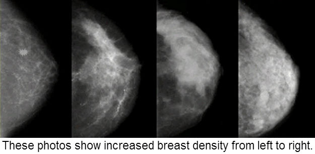
A major drawback to establishing broad screening guidelines is trying to make one size fit all within large populations. Now, in a just-published study in the Annals of Internal Medicine, researchers from the Cancer Intervention and Surveillance Modeling Network, collaborating with the Breast Cancer Surveillance Consortium (BCSC), suggest that for women ages 50 to 75, screening intervals should be based on a combination of breast density (a radiological finding on the mammogram, seen in the images at the top of this blog) and a woman's other risk factors for breast cancer (such as a family history), an approach that may be better tailored to each individual.
The authors of this study recommend that, for women with low breast density on mammograms and low to average risk factors for breast cancer, screening every 3 years may be sufficient, while for women with radiologically-dense breasts and/or other risk factors, yearly screening should be undertaken. 
Having radiologically-dense breast tissue on mammograms has been linked to an increased risk of developing breast cancer coupled with greater difficulty in detecting tumors when they are small because the dense breast tissue often obscures them.
That said, what else besides a family history puts a woman in the high-risk category?
  • ​Obesity (greater than 20% over ideal body weight)
  • Enlarged thyroid (including Hashimoto’s thyroiditis)
  • Diabetes
  • Never having been pregnant (nulliparity)
  • First pregnancy age 32+
  • Regular alcohol (all types) consumption starting in teenage years
  • Smoking, including chronic exposure to second-hand smoke
  • Prolonged use of oral contraceptives during reproductive years
  • Hormone replacement therapy (HRT) after menopause
Taking into account this information, if one or more items in this list applies to you, then irrespective of breast density on a mammogram, your risk for developing breast cancer is significantly higher than average and I suggest you opt for yearly screening mammograms rather than every three years! 


0 Comments

July 30, 2016

7/30/2016

0 Comments

 
In a just-published article, statnews.com science writer, Sharon Begley, reviews the history of failure of Alzheimer's drugs in clinical trials. The latest casualty, a drug called LMTX that targets a prion-like protein, called tau, bitterly disappointed the research community. In a 2012 blog I wrote for CTV.ca, I reviewed the pathology of Alzheimer's Disease (AD), discussed the controversy surrounding the relevance of tau and  another prion-like protein, beta-amyloid, and reported on exciting rodent research showing that tau spreads through the central nervous system like an infection, affecting the same brain memory centres in mice that are involved in humans with AD . Commenting on the study, one expert suggested that, to be effective, treatment for Alzheimer's must target both tau and beta-amyloid. Although a drug called PBT2 that I mentioned at the end of my article also subsequently failed in a phase 3 study, I remain hopeful that just-announced trials of  nilotinib (Tasigna), already in clinical use for the treatment of chronic myeloid leukemia, may turn the corner on both Parkinson's and Alzheimer's therapy. Time will tell...
Picture

New discoveries raise hope for more effective Alzheimer's treatment
by Dr. Lorne Brandes  February 15, 2012 
 
"The ‘Big C’ has to be the worst of all diseases,” many people comment when they learn than I am an oncologist.
My answer often surprises them: “I would rather take my chances with cancer than be diagnosed with Alzheimer’s.”
My reasoning? A diagnosis of early cancer still leaves lots of room for hope; our treatments are increasingly effective and commonly lead to complete recovery. Even in its advanced, incurable stages, many cancers can be palliated for months or, in some cases, years. Even at the end of the road, the mind usually remains intact, allowing people to reminisce about the good times, mend broken fences, and say their final goodbyes to friends and loved ones.
Alzheimer’s dementia (AD) is quite another story. Since it was first described in 1906 by Dr. Alois Alzheimer, a German psychiatrist and neuropathologist, this ever-increasing degenerative brain disease has remained poorly understood. Despite the recent availability of drugs, such as donepezil (Aricept), that can sometimes slow down early AD for a few months, there remains no effective treatment to prevent its relentless progression.
As a result, to be diagnosed with Alzheimer’s, even at an early stage, currently leaves little, if any, room for optimism or hope. What can be worse than the uncomprehending, blank stare of a previously vital human being whose memories of people, places and events have been progressively erased over a relatively short time?
But now, two newly-published papers suggest that we may be closer than previously believed to understanding what goes wrong in the brains of Alzheimer’s patients. As a result, we now have promising new leads on how to stop the disease in its tracks. Indeed, one novel therapy, based on a concept reported in one of the papers, has already shown promise in an early (phase 2) clinical trial, with a follow-up study now underway.
To understand these new discoveries, let us first review what is known about two abnormal structures, called plaques and tangles, observed under the microscope in brain tissue of patients with AD.
Plaques (also called “senile plaques”) are made up of clumps of a structurally abnormal (misfolded) protein, called beta amyloid. Thin fibres of this substance first accumulate around nerve cells in a vital memory area of the brain, called the entorhinal cortex.
Eventually, smaller, soluble beta amyloid molecules, called “ toxic oligomers”, break away from the plaques. This dissolvable form of beta amyloid is thought to interfere with chemicals (called neurotransmitters) through which the brain sends out signals over nerve networks.
Beta amyloid is derived from “ amyloid precursor protein” (APP), made by a gene on chromosome no. 21. Why is this important to know? Because patients with Down syndrome, resulting from an extra chromosome 21, all develop AD if they live long enough. That is probably the most persuasive reason for linking AD with an abnormal buildup of beta amyloid in the brain.
Tangles (also called “ neurofibrillary tangles”) form the second piece of the Alzheimer puzzle. We now know that, like plaques, thin spaghetti-like tangles are also made up of clumps of an abnormally folded protein, in this case, a substance called “tau”.
Whereas beta amyloid plaques accumulate outside nerve cells, toxic amounts of tau accumulate inside, where they disrupt a tubular canal system (called microtubules) that serve as conduits for vital nutrients and chemicals that help nerve cells function and remain healthy. In addition, tau also increases the toxic effects of a second cell-damaging protein, called alpha (α)-synuclein , implicated in many neurodegenerative diseases, including AD, Parkinson’s and multiple sclerosis.
While recent studies suggest that beta amyloid drives the formation of tau, a fierce debate continues over whether either of these misfolded proteins actually cause the disease.
Indeed, leading Alzheimer researcher, Dr. Rudy Castellani, believes that the beta amyloid hypothesis is “deeply flawed and certainly unproven.” He, and others, have suggested that beta amyloid is irrelevant, simply part of the flotsam and jetsam resulting from cell damage caused by some yet undiscovered cause.
Calling for a fresh start, Castellani says , “I think we have to throw the kitchen sink at the problem. Everything should be on the table, including a poly-therapy approach that encompasses multiple constructs and hypotheses.” But, although his discouragement over the lack of progress is understandable, the two new studies, to which I alluded earlier, give fresh insight into why both beta amyloid and tau may be important factors in AD.
The first , led by scientists Karen Duff and Scott Small of New York’s Columbia University, used mice that were genetically-engineered to specifically produce abnormal human tau protein in cells in the entorhinal cortex. Over a period of several months, the aberrant tau protein was found to spread “ like an infection” along nerve networks connecting the entorhinal cortex to other memory centres of the brain, a process that closely mimics the progression of AD in humans.
Prior to this experiment, doctors wondered if AD starts in the entorhinal cortex because its cells are somehow the most vulnerable to beta amyloid damage. According to this hypothesis, the disease eventually affects memory cells in other “bad neighborhoods” in the brain where beta amyloid deposits occur.
But now, says Alzheimer expert, Dr. John Hardy of London’s University College, “with the [new] mouse studies, the issue of a bad neighborhood is settled….It isn’t a bad neighborhood. It is contagion from one neuron to another…[and] if tau spreads from neuron to neuron, it may be necessary to block both beta amyloid production, which seems to get the disease going, and the spread of tau, which continues it, to bring Alzheimer’s to a halt.”
One way to prevent the spread of tau is to develop a specific antibody that, by latching on to the abnormal protein, arms the immune system to remove it; another is to identify membrane-blocking drugs that prevent tau from leaving cells so that it can not travel along nerve networks.
As for blocking beta amyloid, therapies that reduce its level in the brain have been, so far, unsuccessful in reversing dementia, a fact duly noted by Dr. Castellani and other critics of the beta amyloid hypothesis.
However, skepticism about the value of attacking beta amyloid may be about to change as a result of the second new study, published by a research team led by Dr. Susan Lindquist of the Whitehead Institute for Biomedical Research in Cambridge, MA. Lindquist’s group is interested in trace metals, such as iron, copper and zinc, that are required by enzymes and neurotransmitters to function normally. They have identified a family of compounds, called 8-hydroxyquinolines (8-OHQ), that protect yeast cells from TDP-43, a metal-binding neurotoxic protein implicated in amyotrophic lateral sclerosis (ALS) that appears to behave like beta amyloid.
One 8-OHQ derivative mentioned in Lindquist’s study is PBT2, an AD drug currently being tested by Prana Biotechnology, a small Australian drug company. According to Prana, PBT2 specifically blocks the ability of soluble beta amyloid to tie up copper and zinc in the small spaces (called synapses) between nerve endings, an effect they believe explains the significant improvement in cognitive function observed in an earlier phase 2 study.
“Unlike other [beta amyloid] strategies, we target the initial disease progression steps that result in A‐beta becoming toxic, [that] being the interaction with metals in the brain… PBT2 targets these metals, restoring neuronal function to treat the disease,” the company states.
Now, with a new trial of PBT2 finally underway, that hypothesis will be put to the test. Needless to say, the study and its outcome will be followed with great interest. 
0 Comments

July 28, 2016

7/28/2016

0 Comments

 
For some years, scientists have argued over whether a chemical, called 4-methylimidazole (4-MI), that gives colas their characteristic caramel color, poses a cancer risk to humans. The latest example is a study, published in 2015 by researchers at Johns Hopkins in conjunction with US Consumer Reports, that called for action to reduce the amount of 4-MI in various soft drinks. Once more there was a swift rebuttal from industry; moreover, the FDA, which has been studying 4-MI for several years, does not believe that there is yet enough scientific evidence on which to act, so the argument continues to go round and round. Here is an examination of the issue that I posted on CTV.ca/health in 2011.
Picture

​Does the colour in cola cause cancer? A reality check
by Dr. Lorne Brandes  February 23, 2011 
​

First, I must make the following disclosure: I can’t remember the last time I drank a Coke, Pepsi or any other brand of cola. If it had to depend on me, the soft-drink industry would not exist.
Second, I must share a fond, but quirky, memory with you. Back in my medical school days, I had a wonderful professor of dermatology, the late Dr. William (Bill) Pace, who introduced my wife and me to his favorite dessert, a delicacy his family unappetizingly dubbed “boiled can”.
Its name was derived, literally, from boiling a small unopened can of Carnation condensed milk in a pot of water for a precise number of minutes. After cooling the can to room temperature, the lid was opened and the container overturned on a plate. Out plopped a light brown-coloured pudding with an out-of-this-world taste.
“Boiling turns the sugar in the milk to caramel, but you mustn’t overboil it or the whole thing will turn dark and bitter,” Bill explained.
Such was my unexpected introduction to the process of caramelization, a chemical reaction that results when various types of sugar (fructose and glucose, for example) are heated to form caramel, the substance that imparts its distinctive, pleasantly sweet flavour to a wide assortment of food, candy and beverages.
In addition, caramel adds colour, ranging from gold to brown, to a wide range of products, from colas to Worcestershire sauce, coffee, wine and baked goods. However, to meet the many different uses of caramel by the food industry, its properties are manipulated by heating sugars with chemical additives, such as ammonia and sulfite, to form what are called “E150 caramels”.
For example, colas require negatively-charged caramel molecules, generated when ammonia is added to the sugar; otherwise, if positively charged, the caramel would react with phosphoric acid (the substance that gives colas their “tang”) and precipitate out of solution, resulting in a loss of the brown colour.
However, heating sugar in the presence of ammonia generates many other compounds in addition to caramel; among them are by-products called “methylimidazoles”.
Rodent studies carried out in 2008 by scientists at the U.S. Government’s National Institute of Environmental Health Sciences revealed that a two-year exposure to increasing concentrations of 4-methylimidazole (4-MI), present in colas, caused a significant increase in a type of lung cancer, called alveolar/bronchiolar carcinoma, in both male and female mice, but not in rats. There was a non-significant trend towards an increase in a type of leukemia in female rats, but no evidence of carcinogenicity in male rats.
Based on these findings, in 2009 the state of California posted its intention to list 4-MI as a carcinogen under the Safe Drinking Water and Toxic Enforcement Act of 1986 (Proposition 65).
Now, following California’s lead, a Washington, DC organization, called the Center for Science in the Public Interest (CSPI), has just petitioned the Food and Drug Administration (FDA) to “revoke regulations authorizing the use [of] caramel colorings that are produced by means of an ammonia or ammonia-sulfite process and contain 2-methylimidazole and 4-methylimidazole, both of which are carcinogenic in animal studies.”
"If consumers want another reason to avoid soda pop, this is a good one. It makes no sense to leave these in the food supply," the group's Executive Director, Michael F. Jacobson, commented.
The response from the beverage industry was swift: "The safety of our products is the foremost priority for our companies. Consumers can take confidence in the fact that people have been safely drinking colas for more than a century, as well as consuming the wide variety of foods and beverages containing 4-MI, from baked goods and breads to wine and coffee. 4-MI is found in trace amounts in a wide variety of foods and beverages, including Coca-Cola. In fact, it forms normally in the 'browning reaction' while cooking, even in one's own kitchen."
As for the FDA, in answer to an inquiry about the CSPI petition from a blog site called Consumer Ally, the agency noted that “its assessment [of the data] will dictate what, if any, regulatory action needs to be taken.” According to the website, in making this statement, the FDA acknowledged that 4-MI is on a list of chemicals to be reviewed for carcinogenicity by the World Health Organization’s International Agency for Research on Cancer.
What is the truth here? Clearly, 4-MI, a by-product of caramelization in the presence of ammonia, is a carcinogen in the mouse, but not in the rat. As for humans, who have been “safely drinking colas for more than a century”, who knows? There are no human studies of 4-MI from which to remotely glean an answer.
So, will the FDA act to remove the ammonia/caramel colouring from colas? Don’t bet on it.
The reality is that any cancer danger from non-diet Coke and Pepsi arises far less from their small content of 4-MI than from their astronomically high content of sugar. Published studies show that the daily consumption of sugar-rich soft drinks, colas among them, contribute significantly to the epidemic of obesity in our society. And make no mistake about it, up to half of all cancers, whether in rodents or humans, are directly related to obesity, and may be prevented by staying slim and trim.

0 Comments

July 19, 2016

7/19/2016

0 Comments

 
Despite the Cancer Moonshot initiative spearheaded by VP Joe Biden, there is still no shortage of people who believe that a cure for cancer has already been found but is being suppressed by the multi-billion dollar "Cancer Industry". When it comes to the conspiracy theorists, nothing seems to change their minds...witness a blog I posted for CTV.ca/health in 2009.​
Picture
​
​Is there a conspiracy to suppress a cure for cancer?
by Dr. Lorne Brandes  November 11, 2009 

Do you remember the movie “Conspiracy Theory”? It was about a New York taxi driver who witnessed life through paranoid-coloured lenses. Except, as it turned out, a government conspiracy to “get him” really was lurking in the wings.
Yet, do not believe for a moment that “conspiracy theorists” are just a Hollywood invention. They also exist in real life. For example, there are people who believe that drug companies don’t want to find a cure for cancer.
As proof, you need look no further than two comments posted online in response to one of my blogs describing the hard realities of cancer drug development.
“Why would the Pharma companies want a cure for cancer? Their bottom line would suffer ... It’s all about money to them…” wrote someone calling himself “Steve in Toronto”.
And he isn’t alone. Linda from the UK added, “….[Steve's] dead right about the pharmaceutical companies. It's a sad state of affairs, but it's true. Money, money, money.”
While most of you may disagree with Steve and Linda, I suspect that more than a few believe, as they do, that curing cancer is not in the financial interest of big pharma or, for that matter, of anyone in the “cancer establishment”, including oncologists such as me.
And quite frankly, it is all too easy to be suspicious of big pharma. Good behaviour is not the drug industry’s strong suit.
Just recently, Pfizer, was slapped with a $2.3 billion fine by the U.S. Justice Department for the fraudulent “off-label” promotion of some of its major drugs. This is the fourth time since 2002 that Pfizer , or one of its subsidiaries, has been fined over illegal marketing practices. As a result, one might be justified in wondering what other deceptions lurk in the shadows of the corporate boardroom.
Drug companies aside, is there a high-level conspiracy to suppress a cancer cure? One only has to surf the internet to see the depth of conviction, even among some notable “experts”, that there is.
For example, the late Dr. Robert Atkins, of high-fat, low-carbohydrate diet fame, was quoted on one website as saying, “There is not one, but many cures for cancer available. But they are all being suppressed by the ACS (American Cancer Society), the NCI (National Cancer Institute) and the major oncology centres. They have too much interest in the status quo.”
Truth be told, at the same time as he was impugning the ACS and NCI, Dr. Atkins was pushing his own “model of cancer treatment” that included his diet, and a variety of unproven or disproven substances such as laetrile, 714-X and essiac.
On the other hand, after examining all the facts, Michael Higgins, an Australian engineer afflicted with cancer, came to a very different conclusion.
“I know there isn't any cancer conspiracy because I know that the people doing and running the research are human,” he wrote. “Their lives, like mine, have been touched by cancer. They, like me, would do anything to save the lives of the people they love. Furthermore, I assume that any treatments associating themselves with a conspiracy theory have something to hide—the simple fact that [they don’t] work.”
But let’s face it. No matter what anyone says, cancer conspiracy advocates will not be swayed from the belief that, to insure their own survival, the “cancer treatment establishment” in general, and the pharmaceutical industry in particular, is not interested in curing cancer.
While I, myself, have strongly criticized the exorbitantly high prices that pharmaceutical companies command for new, often marginally-effective, anti-cancer drugs, and have described their reluctance, for financial reasons, to develop new uses for old drugs that might benefit cancer patients, I do not believe for one minute that they, let alone anyone else in the “establishment”, are suppressing anything.
How can I be so sure?
As the late Michael Higgins so eloquently observed, few have been untouched by this terrible disease. That’s reason in itself.
So if there exists a conspiracy among all those involved, however imperfectly, in the war against cancer, it is simply this: to find the cure. Period.


0 Comments

July 14, 2016

7/14/2016

0 Comments

 
Last October, neuroscientists at Washington's Georgetown University (GU) made headlines with a small study in 12 patients with advanced Parkinson's Disease (PD), including PD with Lewy Body Dementia, eleven of whom responded rather dramatically to a low oral daily dose of the chronic myeloid leukemia drug, nilotinib (Tasigna). When the drug was stopped, the patients' conditions returned to baseline.
Now, in a press release coinciding with publication of that study in the Journal of Parkinson's Disease, the GU investigators have announced the launch of 2 new placebo-controlled phase II trials of nilotinib in patients with PD/Lewy Body Dementia and Alzheimer's Dementia (AD). A small companion study testing nilotinib in patients with amyotrophic lateral sclerosis (ALS) will also be conducted.
As one who has a great deal of interest in the science behind this approach, I am reprinting the GU press release. We all wish for a successful outcome of this potentially ground-breaking therapy!

WASHINGTON (July 11, 2016) — A small phase I study provides molecular evidence that an FDA-approved drug for leukemia significantly increased brain dopamine and reduced toxic proteins linked to disease progression in patients with Parkinson’s disease or dementia with Lewy bodies. Dopamine is the brain chemical (neurotransmitter) lost as a result of death of dopamine-producing neurons in these neurodegenerative diseases.
Researchers from Georgetown University Medical Center (GUMC), say the findings, described in the Journal of Parkinson’s Disease, support improved clinical outcomes observed and first reported at the Society for Neuroscience annual meeting in October 2015.
The study tested nilotinib taken daily for six months. A much smaller dose of nilotinib (150 or 300 mg once daily) was used compared to the dose for chronic myelogenous leukemia (300-400 mg twice daily). Twelve patients were enrolled in the clinical trial — one patient withdrew due to an adverse event. Researchers say the drug appears to be safe and well tolerated in the remaining 11 participants who completed the study.
In addition to safety, the researchers also examined biological markers in the blood and cerebral spinal fluid as well as cognitive, motor and non-motor improvement. They found significant signs that nilotinib may provide benefit for patients with these neurodegenerative diseases.
“These results need to be viewed with caution and further validated in larger placebo controlled trials, because this study was small, the patients were very different from each other, and there was no placebo,” says the study’s senior investigator, Charbel Moussa, MD, PhD, scientific and clinical research director of the GUMC Translational Neurotherpeutics Program.

Among the biomarker findings were that:

(1) The level of the dopamine metabolite homovanillic acid — an indicator that dopamine is being produced — steadily doubled, even with the loss of most dopamine neurons. Most study participants were able to stop using, or reduce their use of, dopamine replacement therapies;
(2) 
The level of the Parkinson's related oxidative stress marker DJ-1 — an indicator that dopamine-producing neurons are dying — was reduced more than 50 percent after niltonib treatment; and
(3) 
The levels of cell death markers (NSE, S100B and tau) were significantly reduced in cerebrospinal fluid (CSF) suggesting reduced neuronal cell death.
In addition, Moussa adds that it appears nilotinib attenuated the loss of CSF alpha-synuclein, a toxic protein that accumulates within neurons, resulting in reduced CSF levels in both Parkinson’s disease and dementia with Lewy bodies.

The researchers also said that all 11 patients who tolerated the drug reported meaningful clinical improvements. All patients were at mid-advanced stages of Parkinsonism and they all had mild to severe cognitive impairment.
“Patients progressively improved in motor and cognitive functions as long as they were on the drug — despite the decreased use of dopamine replacement therapies in those participants with Parkinson’s and dementia with Lewy bodies,” says the study’s lead author, Fernando Pagan, MD, medical director of the GUMC Translational Neurotherpeutics Program and director of the Movement Disorders Program at MedStar Georgetown University Hospital.
But three months after withdrawal of the drug, participants returned to the same reduced cognitive and motor state they had before the study began, Pagan adds.
Some serious side effects were reported including one patient who withdrew at week four of treatment due to heart attack and three incidents of urinary tract infection or pneumonia. The researchers say these incidents are not uncommon in this patient population, and additional studies are needed to determine if the adverse events are related to use of nilotinib.
“Long term safety of nilotinib is a priority, so it is important that further studies be conducted to determine the safest and most effective dose in Parkinson’s, says Pagan.
The researchers designed the clinical trial to translate several notable observations in the laboratory. The preclinical studies, led by Moussa, showed that nilotinib, a tyrosine kinase inhibitor, effectively penetrates the blood-brain barrier and destroys toxic proteins that build up in Parkinson’s disease and dementia by turning on the “garbage disposal machinery” inside neurons.
Their published studies also showed nilotinib increases the levels of the dopamine neurotransmitter — the chemical lost as a result of neuronal destruction due to toxic protein accumulation — and improves motor and cognitive outcomes in Parkinson’s and Alzheimer’s disease animal models.
“Our hope is to clarify the benefits of nilotinib to patients in a much larger and well controlled study. This was a very promising start,” Moussa says. “If these data hold out in further studies, nilotinib would be the most important treatment for Parkinsonism since the discovery of Levodopa almost 50 years ago.”  
He adds, “Additionally, if we can validate nilotinib effects on cognition in upcoming larger and placebo controlled trials, this drug could become one of the first treatments for dementia with Lewy bodies, which has no cure, and possibly other dementias.”
Two randomized, placebo-controlled phase II clinical trials are planned for summer/fall in Parkinson’s and Alzheimer’s diseases. The Translational Neurotherpeutics Program is also planning a small trial in ALS (Lou Gherig’s disease).
According to Novartis, the cost (as of Oct. 2015) of nilotinib for the treatment of CML was about $10,360 a month for 800 mg daily. The dose used in this study was lower —  150 and 300 mg daily.
The phase I study received philanthropic funding and was supported by the Georgetown-Howard Universities Center for Clinical and Translational Science.
Moussa is listed as an inventor on a patent application that Georgetown University filed related to the use of nilotinib and other tyrosine kinase inhibitors for the treatment of neurodegenerative diseases. 
0 Comments

July 6, 2016

7/6/2016

0 Comments

 
Vitamin supplements continue to be a multi-billion dollar industry despite the fact that multiple large population studies have failed to show any benefit to health in well-nourished individuals. Indeed, people who consume daily high doses of some vitamins may increase their risk of certain cancers. For example, in two separate studies, smokers who took daily beta carotene supplements had a higher risk of developing lung cancer than smokers who did not.  
Another supplement that has been linked to increased cancer risk is folic acid. On the other hand, naturally-occurring folate in food may be beneficial. Here is a CTV.ca/health blog I posted in 2009 that reported on an increased risk of prostate cancer in men who took folic acid supplements.
Picture

Folic acid and prostate cancer: Too much of a good thing?
by Dr. Lorne Brandes  March 13, 2009 
 
This week brings yet another cancer study pointing to the potential danger of a vitamin supplement. This time it's folic acid, a synthetic form of folate, one of the essential B-complex vitamins that naturally occurs in green leafy vegetables (the word folate is derived from folium, Latin for "leaf").
In addition to playing a vital role in the production of our cells' DNA, folate is essential for the normal development of the nervous system in the fetus and also for preventing anemia. Short and simple, folate is necessary for life.
Now for the bad news.
In a paper just published in the Journal of the National Cancer Institute, researchers from the University of Southern California studied the development of prostate cancer in 643 men who were randomized to take a daily placebo pill or a daily 1 mg folic acid pill.
Here's the story on CTV.ca: Folic acid linked to higher risk of prostate cancer
Somewhat shockingly, after 10 years of follow-up, those who took folic acid had a prostate cancer rate 2.6 times higher than those who took the placebo!
It should be pointed out that the prostate cancer study was actually an extension of another published study, conducted between 1994 and 2006, called the Aspirin/Folate Polyp Prevention Study (AFPP). That study, published in the Journal of the American Medical Association, found that while aspirin reduced the risk of colon polyps, folic acid appeared to increase the risk of advanced and multiple polyps.
Should we be concerned by these findings? No and yes.
No, because the current study is rather small. As a result, its conclusion that folic acid supplements may be associated with an increased rate of prostate cancer could be due to chance alone. Indeed, blood tests in the two groups of men showed no significant difference in folate levels to account for the observed increase in prostate cancer in men who took folic acid.
Yes, because in the 1940s, Dr. Sidney Farber observed that giving folic acid to children with acute leukemia actually made the disease worse. He reasoned that, since folate increases the production of DNA, it may actually have caused the leukemia cells to divide faster.
Acting on Farber's hunch, his colleague, Dr. Y. Subbarao, Research Director at Lederle Laboratories, soon formulated an anti-folate drug, called methotrexate, that prevents the vitamin from building DNA. The result? Not only was methotrexate the first effective drug treatment for acute leukemia, it is still a mainstay in the treatment of many cancers!
Clearly, then, folate appears to be a double-edged sword: not enough and health suffers; too much and adverse effects, such as increased cell division, possibly resulting in cancer, may result.
The story is also complicated by the fact that folic acid as a supplement may fundamentally differ from natural folate in its biological effects. For example, studies suggest that folate (as opposed to folic acid) may actually protect against stomach and pancreatic cancer.
Yet, because cereals and other grains are now being fortified with folic acid, it is becoming increasingly difficult to separate naturally-occurring folate from the synthetic form in many of the foods we eat. As a result, and because of the studies reported here, I would be concerned that too much fortification could tip the balance from benefit to risk.
That aside, once again the bottom line is this: get your vitamins naturally, in the good food you eat, rather than in a bottle from the health food store. With each new published study comes increasing evidence that, as opposed to a healthy diet rich in fruits and vegetables, most vitamin and mineral supplements do you no additional good and, in some cases, may cause harm.
 
 
0 Comments

June 28, 2016

6/28/2016

0 Comments

 
An important study just published in the Annals of Internal Medicine has found no evidence that receiving a blood transfusion from donors with a neurodegenerative disease, such as Alzheimer's or Parkinson's, increases the risk for neurodegenerative disease in the recipient. This finding may go some distance in allaying concerns that these diseases can be spread from person to person. Underlying this worry has been scientific evidence that abnormal (misfolded) proteins, similar to prions, are the likely cause of many neurodegenerative disorders that spread through the central nervous system over time.
That said, did you know that prion-like proteins, implicated in dementia, also appear to be vital for the formation and retention of normal memory? Read more in this re-posted CTV.ca/health blog I wrote in 2012.
Picture

​
​The narrow line between forming memories and losing them

 by Dr. Lorne Brandes   Jul. 11, 2012 

Judging by the number of “recommenders” on the Facebook link, a lot of readers were interested in last week’s blog, Targeting iron in Parkinson’s Disease (PD). Apart from the role that iron may play in neurodegenerative diseases, perhaps what fascinated most people is the likelihood that a prion-like protein, called alpha-synuclein , may initiate Parkinson’s and then spread it from one brain area to another.
To review,  prions (derived from “protein infection”) can exist in two forms, each exhibiting a distinct shape and behavior:
  • in their normal (or “recessive”) form, prions have a corkscrew shape, called an alpha-helix; recessive prions are manufactured by our genes, are the most common type, and reside harmlessly in cells.
  • however, when, for a variety of reasons, several recessive prions cluster together to form oligomers [from the Greek “oligo”, meaning “a few”], their shape changes into a misfolded (“dominant”) protein that resembles a pleated sheet.
With the change in shape comes a change in character: unlike their recessive cousins, dominant prions self-replicate, using themselves as a template to form new dominant prions. For poorly-understood reasons, they can also behave as rogues, killing brain cells as they spread, like an infection, through the nervous system.
Now, with the identification of neurotoxic prion-like proteins, such as alpha-synuclein in PD, and beta-amyloid and tau in Alzheimer’s dementia (AD), scientists are openly discussing the hypothesis that many common human neurodegenerative diseases are “ prion disorders ”, not unlike mad cow disease (BSE) in cattle and scrapie in sheep.
But trumping even that possibility is the amazing discovery that those very same dominant prion oligomers are essential for the formation of long-term memory!
The earliest indication that this might be so surfaced over a decade ago in the Columbia University laboratory of Dr. Eric Kandel, who shared the 2000 Nobel Prize in Medicine for his pioneering work in brain signaling (more specifically, how the brain responds to environmental stimuli and, in the process, forms and retains memories).
While experimenting on the sea slug (Aplysia), a model of nerve function developed by Kandel, Dr. Kausic Si, a postdoctoral fellow, discovered that a vital protein, called CPEB , continuously replenished other proteins that maintain and enhance the function of new nerve endings, or synapses , that sprout in response to operant conditioning (learning), or to repeated exposure to serotonin , a brain neurotransmitter. 
That of itself was an important new discovery. Yet, both Si and Kandel knew that the life of most proteins is short, usually hours. How was CPEB, itself a protein, able to persist and sustain the new nerve endings for long periods of time, perhaps for the life of the sea slug?
After analyzing its structure, Si came up with a startling finding. Kandel describes what happened next in his must-read book, “ In Search of Memory ”:
“I remember it was a beautiful New York afternoon in the spring of 2001, the bright sunlight rippling off the Hudson River outside my office windows, when Kausik walked into my office and asked, ‘What would you say if I told you that CPEB has prion-like properties?’
“A wild idea! But if true, it could explain how long-term memory is maintained in synapses… Clearly, a self-perpetuating molecule could remain at a synapse indefinitely, regulating the local protein synthesis needed to maintain newly grown synaptic terminals.”
Further experiments conducted in yeast cells by Si cemented the observation; CPEB, did indeed have dominant prion-like properties.
But it was one thing to show that CPEB resembled a dominant prion and another to establish conclusively that this likeness was essential to forming and maintaining long-term memory.
That proof came earlier this year. In a study published in Cell, scientists led by Dr. Si, now at the Stowers Institute for Medical Research in Kansas City, showed that, like CPEB in the sea slug, Orb2, a similar nerve cell protein in fruit flies, looks and behaves like a dominant prion oligomer.
Once that was confirmed, Si and his colleagues carried out two operant conditioning experiments in male fruit flies: learning to avoid a female mated to another male; and learning to associate a specific odour with a food (sugar) reward. The result? Orb2 conferred long-term avoidance and association memory (lasting beyond 48 hours) in normal fruit flies. In contrast, mutant fruit flies that carried only a recessive (non-replicating) form of Orb2, retained what they had learned for 24 hours and then completely forgot when retested at 48 hours.
"Self-sustaining populations of [dominant prion oligomers] located at synapses may be the key to the long-term….changes that underlie memory; in fact, our finding hints that oligomers play a wider role in the brain than has been thought," Dr. Si told Science Daily.
He and his team now plan to see how long Orb2 must be present to keep a memory alive. "We suspect that [it] need[s] to be continuously present, because [it is] self-sustaining in a way that [recessive] Orb2 [in mutant fruit flies] is not," Si commented .
As Dr. Kandel notes in his book : “…basic science can be like a good mystery novel with surprising twists: some new, astonishing process lurks in an undiscovered corner of life and is later found to have wide significance. This particular finding [that CPEB is prion-like] was unusual in that the molecular processes underlying a group of strange brain diseases are now seen also to underlie long-term memory, a fundamental aspect of healthy brain function. Usually, basic biology contributes to our understanding of disease states, not vice versa.”
By now, it should be apparent that there is a fine line between forming and losing memory: in normal circumstances, dominant prions giveth; in pathological circumstances, they taketh away.
An answer to the riddle of what goes awry to make them “taketh away” could allow us to prevent many, if not all, neurodegenerative diseases.
0 Comments

 June 27, 2016

6/27/2016

0 Comments

 
Thanks to oncologists at Houston's MD Anderson Cancer Center, we now know how, and when, to treat breast cancer in pregnancy. Here is a re-posting of a blog on the subject that I wrote for CTV.ca/health in 2010. The information remains pertinent in 2016.
Picture


Treating breast cancer in pregnancy: Good news for mother and child
by Dr. Lorne Brandes   October 22, 2010 

​For decades, it was every oncologist’s nightmare: a pregnant woman diagnosed with rapidly growing breast cancer. What to do? Terminate the pregnancy? Wait until delivery before starting chemotherapy? Treat immediately but reduce the dose of chemotherapy in an attempt to protect the fetus? Until recently, nobody knew the answers to these very difficult questions.
Now, thanks to pioneering studies carried out at Houston’s prestigious University of Texas MD Anderson Cancer Center, we do. Not only has a clear roadmap emerged to help us safely navigate this minefield, but we can also confidently offer a much more encouraging prognosis to the pregnant patient and her unborn baby. Here is the story.
Starting in 1992, MD Anderson oncologists began treating pregnant women with standard (full) doses of three widely-used breast cancer drugs in a regimen called "FAC" (5-fluorouracil, doxorubicin [Adriamycin] and cyclophosphamide). To minimize harm to the fetus, they waited until after the first trimester to begin the chemotherapy. After delivery, radiation treatment, and additional drugs, such as tamoxifen and Herceptin, were administered when indicated.
Close follow-up was provided over the ensuing years to monitor the mothers for any signs of recurrence and to assess the health and development of the children.
The first encouraging news emerged in a 2006 paper that described the MD Anderson team’s results in the first 57 women and their offspring. Not only was the short-term outcome better than expected, with 44 of 57 (77%) women alive and cancer-free after 3 years of follow-up, 97% of the children were reported to have "normal development compared to siblings or other children." Only three had congenital birth defects: one with Down syndrome, one with a club foot, and one with a relatively common and treatable abnormality (called "reflux") of the ureters that drain urine from the kidneys into the bladder.
That wasn’t all we learned from the study. Prior to that time, it had been generally believed that breast cancer in pregnant women grows and spreads rapidly because of high levels of maternal estrogen; this hormone drives tumour growth in two-thirds of non-pregnant women with the disease. However, meticulous analysis of the pregnant women’s tumours revealed that they were estrogen-driven in only 30% of cases, while 70% were "triple-negative" high grade cancers that typically grow independent of estrogen and carry a poorer than average prognosis. Suddenly, we had an alternative explanation for the aggressive behaviour of breast cancer in pregnant women!
But the surprises didn’t end there. At the recent 2010 Breast Cancer Symposium, the MD Anderson’s Dr. Jennifer Litton reported that 75 pregnant women have now been treated and followed for an average of five years. Given the aggressive nature of breast cancer in pregnancy, their disease-free survival rate remains remarkably high at 74%.
However, what really caught everyone’s attention was how that result compared to the outcome her group obtained using the same "FAC" chemotherapy treatment in 150 non-pregnant women with breast cancer who were closely matched for age, year of diagnosis, and stage of disease. As opposed to pregnant women, non-pregnant women had a significantly lower five-year disease-free survival rate of 56%!
How to explain the difference? According to Dr. Litton, "We are not sure why our pregnant breast cancer patients had better outcomes than those who were not…is there something biological in the milieu of pregnancy that changes the response to chemotherapy?" Finding that answer will be her group’s "research priority", she said.
As an oncologist who treats this disease on a daily basis, I wish her and her colleagues every continuing success. While much longer follow-up will be required to determine the ultimate cancer-free survival of the mothers, as well as any long-term effects of in utero exposure to chemotherapy in their children, thanks to the MD Anderson researchers we are already light years ahead in our ability to advise, reassure, and treat pregnant women with breast cancer.
 

0 Comments

June 22, 2016

6/22/2016

0 Comments

 
With the recent death of Muhammad Ali from complications of Parkinson's Disease (PD), new attention has turned to the causes and treatment of this all-too-common neurodegenerative disorder. As a result, I am re-posting a blog I wrote for CTV.ca/health in 2012 that examined the role of iron buildup in the brain that may promote the formation of an abnormal prion-like protein, called alpha-synuclein, that many neuroscientists now believe to be at the root of PD and may explain the progressive spread of this disease throughout the brain over time. The blog garnered much interest and many citations on other websites. Here it is.
Picture


Targeting iron in Parkinson’s Disease
by Dr. Lorne Brandes  Jul. 4, 2012 

There are some exciting findings afoot to suggest that too much iron in brain tissue is a common finding in neurodegenerative diseases, and that removing it with certain drugs, called chelators, may significantly improve symptoms.
But to help you understand this, I first need to review some additional scientific information.
In a recent blog on Alzheimer’s Disease (AD), I reported on a misfolded protein, called tau. In its normal form, tau has a corkscrew appearance (called an “alpha-helix”). However, when too much tau is made by cells, multiple tau molecules stick together (aggregate). In the process, tau changes shape (misfolds), becoming a pleated structure (called “beta sheets”) that is toxic to nerve cells.
Of great interest, pleated tau structurally resembles a prion, the infectious agent that is best known for causing mad cow disease (BSE, or bovine spongiform encephalopathy). BSE can be transmitted to humans who eat prion-infected meat (the human form of BSE is Creutzfeldt-Jakob disease, a rapidly-progressive neurodegenerative disorder).
Even if tau looks like a prion, does it behave like one ? It appears that it does: researchers have discovered in mice that, similar to the progression of AD in humans, tau first accumulates in a memory area of the brain called the entorhinal cortex and then spreads along nerve networks “like an infection” to progressively involve other memory centres.
How does pleated tau spread? Scientists believe that it uses itself as a template to form replicas that leave the cell and move from nerve to nerve. That said, unlike BSE, so far there is no evidence to suggest that tau can survive outside the nerve cells of the human brain or be transmitted from one person to another.
Yet, transmissible or not, the prion-like structure and “contagious behaviour” of tau has suddenly become a hot topic among neuroscientists who quickly point to another misfolded (pleated) protein, called alpha (α)-synuclein, a neurotoxin that is strongly linked to Parkinson’s Disease (PD).
Under the microscope, nerve cells affected by PD contain rounded inclusions, called Lewy bodies (named after their discoverer, Dr. Frederic Lewy), that are rich in alpha-synuclein. Pathologists have also identified Lewy bodies in human embryonic stem cells that were implanted into brains years earlier in studies to treat PD.
Noting that these transplanted donor cells must have become “infected” with alpha-synuclein from the recipients’ brains, Dr. Stanley Prusiner, who won the 1997 Nobel Prize for discovering the prion, recently suggested that, indeed, PD may be “a prion disorder”!
Is he overstating the case?
Consider this: we now have evidence that, even before PD affects the brain, it starts in the sympathetic nerve fibres that control bowel function. Researchers have found biopsy evidence of alpha-synuclein in gut tissue up to five years before typical brain-related symptoms of tremor and muscle rigidity occur. Indeed, chronic constipation, likely caused by alpha-synuclein damage to sympathetic nerve cells, may be the earliest symptom of PD.
Over time, the disease appears in (spreads to?) the brain, initially involving the brain stem, and then moving to an area of dark-staining brain tissue, called the substantia nigra (SN).
The cells of the SN produce a neurotransmitter chemical, called dopamine, that is vital to normal brain function. When approximately 80% of the dopamine-producing cells die from the effects of alpha-synuclein, the typical tremor and/or rigidity symptoms of PD appear. 
Although a drug called L-dopa can relieve these distressing symptoms by replenishing the lost dopamine, it is by no means a cure. Nerve cells continue to wither from alpha-synuclein as the disease spreads to other areas of the brain and spinal cord.
Now (and thank you for being patient) here is where iron comes into the picture: in addition to alpha-synuclein, Lewy bodies contain abundant iron. In addition, a heavy buildup of iron is found specifically in brain areas affected by PD.
Perhaps most important of all, new studies show that iron not only regulates cell production of alpha-synuclein, but promotes its misfolding into the neurotoxic prion-like pleated form!
Suddenly, many researchers are suggesting that removing the buildup of iron in brain tissue might be one way to control the production (and, possibly, misfolding) of alpha-synuclein, thereby slowing or halting the progression of PD and other neurodegenerative diseases.
Indeed, early clinical trials of an FDA-approved oral iron chelator, called deferiprone, have shown promise to decrease brain iron and relieve symptoms in a related neurodegenerative disease called Friedreich’s Ataxia (spinocerebellar degeneration).
Could deferiprone also benefit patients with PD? Dr. David Dexter of London’s Imperial College hopes to find out. He is the principal investigator of a phase 2 trial involving 36 patients; the study, which opened in February, has already accrued close to half of the required subjects.
If the results look promising, phase 3 studies in PD, as well as new trials in other neurodegenerative diseases where brain iron levels are high, such as Huntington’s  Disease  and Lewy Body Dementia will almost certainly follow.
But as always, there is a note of caution. Even if iron chelation therapy helps patient with PD, it is unlikely to be curative. Nerve cells that have already succumbed will not return. Alpha-synuclein that is already misfolded might continue to act as a template, spreading the disease. Dopamine will still need to be buttressed by drugs like L-dopa. Moreover, deferiprone has some potentially serious side effects that must be weighed against any potential benefits.
Nonetheless, the tantalizing prospect that, through its effects on alpha-synuclein, iron may play a major role in more than one neurodegenerative disease process, and that chelation therapy could be an effective antidote, is worth everyone’s attention
0 Comments

June 21, 2016

6/21/2016

0 Comments

 
Hardly a month goes by without new research findings that may ultimately change our approach to treating breast cancer. Just yesterday, researchers revealed that the cells giving rise to hereditary breast cancer resulting from the BRCA1 mutation can be targeted with an anti-osteoporosis drug called denosumab. This raises the possibility that this monoclonal antibody might actually prevent breast cancer in these women and save them from having to undergo prophylactic mastectomies. However, this approach, although exciting and on solid scientific ground, will need to be carefully assessed in properly-designed clinical trials to know whether it works and is safe over the long term. If so, it will almost certainly become the new approach in this select group of women with a very high lifetime risk of developing breast cancer.
The BRCA1 story reminds me of an important study, called METABRIC, published in 2012, that found there were ten different types of breast cancer. I reported on it in my CTV.ca/health blog, re-posted below.

Picture

I
mproving the treatment of breast cancer: A tale of 10 diseases
by Dr. Lorne Brandes  April 23, 2012 
 
Having treated breast cancer for four decades, I can attest to the progressive improvement in outcome in women diagnosed with early-stage disease. Multi-pronged advances in chemotherapy, radiation and hormonal therapy, aided by earlier detection through regular screening mammograms, have increased the five-year survival rate from 71% when I began practice in the early 1970’s, to 90% in 2012.
Yet, despite this impressive gain, and beyond what the statistics tell us, I am always humbled by our inability to predict the outcome for any specific patient. I remember the faces of those I thought would do well but didn’t, and, conversely, of those I thought might fare poorly but did well.
The reason? Despite the fact that two women may present with breast cancers of identical clinical stage (the degree of spread) and pathological grade (the degree of malignancy under the microscope), their tumours may have radically different biological characteristics and sensitivity to treatment.
For example, approximately two-thirds of malignant breast tumours contain receptors for the female hormones estrogen and/or progesterone. A high content of both hormone receptors generally indicates a better prognosis; recurrence often can be prevented by oral antiestrogen drugs such as tamoxifen or the newer class of aromatase inhibitors. On the other hand, the one-third of tumours that lack hormone receptors are less predictable in their behaviour; nonetheless, chemotherapy significantly improves the outcome in such cases.
Twenty per cent of breast cancers produce high levels of a receptor protein called HER2 . These tumours usually lack estrogen and progesterone receptors, tend to be very aggressive ( high grade) and are more likely to spread (metastasize). However, in recent years, their recurrence rate has been cut in half using chemotherapy in combination with the HER2-targeting drug, Herceptin.
Tumours that lack estrogen, progesterone and HER2 receptors are called triple-negative; they also tend to be high grade. Unfortunately, they often recur despite aggressive chemotherapy and radiation, but those that do not come back within 2 to 3 years after treatment are usually cured.
While tailoring breast cancer therapy according to stage, grade, and receptor status has greatly improved overall survival, choosing the best treatment in each case, based on an accurate prediction of individual outcome, has remained elusive.
But now, that situation is about to change dramatically.
As a result of a landmark study just published online in Nature, we have been given a whole new roadmap of the biology of breast cancer; with it comes the ability to more accurately predict the behaviour of individual tumours.
This breakthrough was made by a consortium (known by the acronym METABRIC) of Canadian- and U.K-based. cancer researchers led by Drs. Samuel Aparicio of Vancouver’s British Columbia Cancer Centre and Carlos Caldas of the Cambridge Research Institute.
By analyzing the DNA in almost 2,000 breast cancer tumours, the scientists discovered that individual tumours contain one of 10 different “clusters” of altered (abnormal) genes. Some of the genes (like the one that produces the HER2 protein) were predictable, but several others had no previously known link to breast cancer.
The group’s conclusion? Breast cancer is not one, but 10 different diseases! Each subtype exhibits unique genetic (and, therefore, biological) characteristics.
But there is more. Because the outcome of treatment was documented in every tumour donor, the prognosis for each of the 10 subtypes of breast cancer is now known!
As one example, tumours with altered “cluster 4” genes, including some high-grade triple-negative cancers, were observed, surprisingly, to be associated with a good prognosis. Intriguingly, cluster 4 alterations do not involve cancer genes; rather, they are associated with immune system genes. This might explain why a rare form of high-grade triple-negative breast cancer, called “medullary”, has a better than average prognosis: medullary breast cancers are typically infiltrated by millions of “killer” immune cells, called CD8 lymphocytes.
These findings raise the possibility that “cluster 4” immune system genes could be harnessed to improve the outcome in other, less responsive forms of breast cancer.
To illustrate, although estrogen receptor-positive breast cancers generally have a good prognosis, the METABRIC study identified two exceptions: an estrogen receptor-positive subtype, containing “cluster 2” gene abnormalities on chromosome 11, responds poorly to blocking estrogen; a second estrogen receptor-positive subtype, containing “cluster 5” abnormalities on chromosome 17 (the HER2 gene), has an extremely poor prognosis despite the use of chemotherapy, Herceptin and antiestrogens.
Could immune stimulation change the poor outcome in these two instances? Evidence that this could be so comes from recent studies showing that chemotherapy given prior to surgery can eradicate breast cancer and significantly increase survival when CD8 cells infiltrate tumours in high numbers.
Commenting on the importance of the METABRIC study, leading British genetics researcher, Professor Charles Swanton, described the new results as an “extraordinary” finding that took our understanding of breast cancer “to the next level”. These findings are “likely to have important implications for clinical trial design in breast cancer and will prime researchers worldwide to define new approaches to treat each subgroup,” he said.
Lead METABRIC researcher, Carlos Caldas, added, “I want to stress, this…wouldn’t have been possible without the breast cancer patients who donated their samples and agreed to take part in the study. None of this would have happened without them, and I’m so grateful for their participation.”
My take? While the full impact of these findings will take years to realize, they are truly revolutionary, certain to ultimately result in more effective breast cancer treatment and improved survival.
 
0 Comments
<<Previous

    Blogs by
    Dr. Lorne Brandes

    Dr. Brandes is a retired oncologist, former CTV.ca blogger, and author of Survival: A Medical Memoir.

    Here are some of his archived CTV blogs.

    Archives

    August 2016
    July 2016
    June 2016
    May 2016

    Categories

    All

    RSS Feed

Powered by Create your own unique website with customizable templates.
  • Home
  • Bio
  • Survival
  • Reviews
  • Blog
  • Media
  • Contact